Bocchetta Martina, Cardoso M Jorge, Cash David M, Ourselin Sebastien, Warren Jason D, Rohrer Jonathan D
Dementia Research Centre, Department of Neurodegenerative Disease, UCL Institute of Neurology, Queen Square, London, UK.
Dementia Research Centre, Department of Neurodegenerative Disease, UCL Institute of Neurology, Queen Square, London, UK; Translational Imaging Group, Centre for Medical Image Computing (CMIC), University College London, UK.
Neuroimage Clin. 2016 Feb 21;11:287-290. doi: 10.1016/j.nicl.2016.02.008. eCollection 2016.
Frontotemporal dementia (FTD) is a heterogeneous neurodegenerative disorder with a strong genetic component. The cerebellum has not traditionally been felt to be involved in FTD but recent research has suggested a potential role.
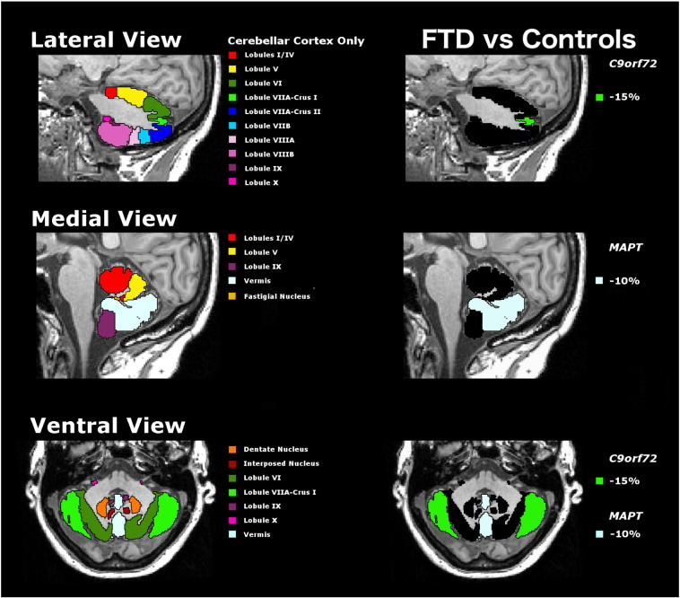
We investigated the volumetry of the cerebellum and its subregions in a cohort of 44 patients with genetic FTD (20 MAPT, 7 GRN, and 17 C9orf72 mutation carriers) compared with 18 cognitively normal controls. All groups were matched for age and gender. On volumetric T1-weighted magnetic resonance brain images we used an atlas propagation and label fusion strategy of the Diedrichsen cerebellar atlas to automatically extract subregions including the cerebellar lobules, the vermis and the deep nuclei.
The global cerebellar volume was significantly smaller in C9orf72 carriers (mean (SD): 99989 (8939) mm(3)) compared with controls (108136 (7407) mm(3)). However, no significant differences were seen in the MAPT and GRN carriers compared with controls (104191 (6491) mm(3) and 107883 (6205) mm(3) respectively). Investigating the individual subregions, C9orf72 carriers had a significantly lower volume than controls in lobule VIIa-Crus I (15% smaller, p < 0.0005), whilst MAPT mutation carriers had a significantly lower vermal volume (10% smaller, p = 0.001) than controls. All cerebellar subregion volumes were preserved in GRN carriers compared with controls.
There appears to be a differential pattern of cerebellar atrophy in the major genetic forms of FTD, being relatively spared in GRN, localized to the lobule VIIa-Crus I in the superior-posterior region of the cerebellum in C9orf72, the area connected via the thalamus to the prefrontal cortex and involved in cognitive function, and localized to the vermis in MAPT, the 'limbic cerebellum' involved in emotional processing.
额颞叶痴呆(FTD)是一种具有强烈遗传成分的异质性神经退行性疾病。传统观点认为小脑不参与FTD,但最近的研究表明其可能发挥作用。
我们调查了44例遗传性FTD患者(20例MAPT、7例GRN和17例C9orf72突变携带者)与18名认知正常对照者的小脑及其亚区域体积。所有组在年龄和性别上进行匹配。在容积性T1加权脑磁共振图像上,我们使用迪德里希森小脑图谱的图谱传播和标签融合策略自动提取包括小脑小叶、蚓部和深部核团在内的亚区域。
与对照组(108136(7407)mm³)相比,C9orf72突变携带者的小脑总体积显著更小(平均(标准差):99989(8939)mm³)。然而,与对照组相比,MAPT和GRN突变携带者未见显著差异(分别为104191(6491)mm³和107883(6205)mm³)。在研究各个亚区域时,C9orf72突变携带者的小叶VIIa - Crus I体积比对照组显著更低(小15%,p < 0.0005),而MAPT突变携带者的蚓部体积比对照组显著更低(小10%,p = 0.001)。与对照组相比,GRN突变携带者的所有小脑亚区域体积均得以保留。
在FTD的主要遗传形式中,小脑萎缩似乎存在差异模式,GRN相对未受影响,C9orf72的小脑萎缩局限于小脑上后部区域的小叶VIIa - Crus I,该区域通过丘脑与前额叶皮质相连并参与认知功能,而MAPT的小脑萎缩局限于蚓部,即参与情绪处理的“边缘小脑”。