The Children's Hospital at Westmead, Psychological Medicine, Locked Bag 4001, Westmead, NSW 2145, Australia; The Brain Dynamics Centre, Westmead Institute for Medical Research, 176 Hawkesbury Rd, Westmead, NSW 2145, Australia; The University of Sydney, Sydney, Australia.
Brain Resource, 235 Jones St, Ultimo, NSW 2017, Australia.
Neuroimage Clin. 2018 Feb 17;18:730-743. doi: 10.1016/j.nicl.2018.02.003. eCollection 2018.
Children and adolescents with functional neurological symptom disorder (FND) present with diverse neurological symptoms not explained by a disease process. Functional neurological symptoms have been conceptualized as somatoform dissociation, a disruption of the brain's intrinsic organization and reversion to a more primitive level of function. We used EEG to investigate neural function and functional brain organization in children/adolescents with FND.
EEG was recorded in the resting eyes-open condition in 57 patients (aged 8.5-18 years) and 57 age- and sex-matched healthy controls. Using a topographical map, EEG power data were quantified for regions of interest that define the default mode network (DMN), salience network, and somatomotor network. Source localization was examined using low-resolution brain electromagnetic tomography (LORETA). The contributions of chronic pain and arousal as moderators of differences in EEG power were also examined.
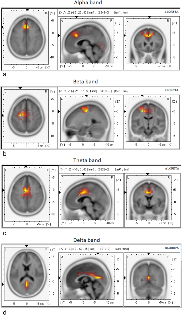
Children/adolescents with FND had excessive theta and delta power in electrode clusters corresponding to the DMN-both anteriorly (dorsomedial prefrontal cortex [dmFPC]) and posteriorly (posterior cingulate cortex [PCC], precuneus, and lateral parietal cortex)-and in the premotor/supplementary motor area (SMA) region. There was a trend toward increased theta and delta power in the salience network. LORETA showed activation across all three networks in all power bands and localized neural sources to the dorsal anterior cingulate cortex/dmPFC, mid cingulate cortex, PCC/precuneus, and SMA. Pain and arousal contributed to slow wave power increases in all three networks.
These findings suggest that children and adolescents with FND are characterized by overactivation of intrinsic resting brain networks involved in threat detection, energy regulation, and preparation for action.
患有功能性神经症状障碍(FND)的儿童和青少年表现出多种无法用疾病过程解释的神经症状。功能性神经症状被概念化为躯体分离,即大脑内在组织的中断和向更原始功能水平的倒退。我们使用脑电图来研究 FND 儿童/青少年的神经功能和功能性大脑组织。
在 57 名患者(年龄 8.5-18 岁)和 57 名年龄和性别匹配的健康对照者中,记录静息睁眼状态下的脑电图。使用地形图,量化定义默认模式网络(DMN)、突显网络和躯体运动网络的感兴趣区域的脑电图功率数据。使用低分辨率脑电磁层析成像(LORETA)检查源定位。还检查了慢性疼痛和唤醒作为 EEG 功率差异的调节剂的作用。
患有 FND 的儿童/青少年在前额(背内侧前额叶皮质[dmFPC])和后部(后扣带回皮层[PCC]、楔前叶和外侧顶叶皮质)的 DMN 以及运动前/辅助运动区(SMA)区域对应电极簇中存在过多的θ和δ功率。突显网络中也存在θ和δ功率增加的趋势。LORETA 在所有三个网络中显示出所有频段的激活,并将神经源定位于背侧前扣带皮层/dmPFC、中扣带皮层、PCC/楔前叶和 SMA。疼痛和唤醒导致所有三个网络中慢波功率增加。
这些发现表明,患有 FND 的儿童和青少年的特征是涉及威胁检测、能量调节和行动准备的内在静息大脑网络过度激活。