University of Central Florida, IST, Modeling and Simulation Department, FL, USA; Semel Institute for Neuroscience and Human Behavior, David Geffen School of Medicine, UCLA, CA, USA.
Imaging Genetics Center, USC Keck School of Medicine, Marina del Rey, CA, USA.
Neuroimage Clin. 2018 Feb 24;18:744-752. doi: 10.1016/j.nicl.2018.02.020. eCollection 2018.
Attention-deficit hyperactive disorder (ADHD) is the most common neurodevelopmental disorder in children. Diagnosis is currently based on behavioral criteria, but magnetic resonance imaging (MRI) of the brain is increasingly used in ADHD research. To date however, MRI studies have provided mixed results in ADHD patients, particularly with respect to the laterality of findings.
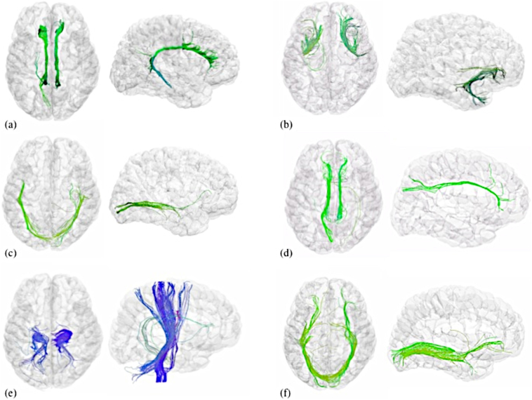
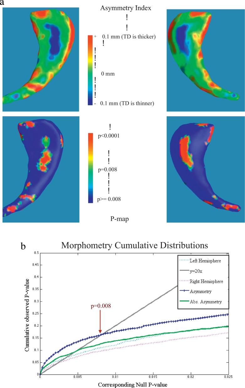
We studied 849 children and adolescents (ages 6-21 y.o.) diagnosed with ADHD ( = 341) and age-matched typically developing (TD) controls with structural brain MRI. We calculated volumetric measures from 34 cortical and 14 non-cortical brain regions per hemisphere, and detailed shape morphometry of subcortical nuclei. Diffusion tensor imaging (DTI) data were collected for a subset of 104 subjects; from these, we calculated mean diffusivity and fractional anisotropy of white matter tracts. Group comparisons were made for within-hemisphere (right/left) and between hemisphere asymmetry indices (AI) for each measure.
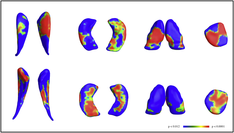
DTI mean diffusivity AI group differences were significant in cingulum, inferior and superior longitudinal fasciculus, and cortico-spinal tracts ( < 0.001) with the effect of stimulant treatment tending to reduce these patterns of asymmetry differences. Gray matter volumes were more asymmetric in medication free ADHD individuals compared to TD in twelve cortical regions and two non-cortical volumes studied ( < 0.05). Morphometric analyses revealed that caudate, hippocampus, thalamus, and amygdala were more asymmetric ( < 0.0001) in ADHD individuals compared to TD, and that asymmetry differences were more significant than lateralized comparisons.
Brain asymmetry measures allow each individual to serve as their own control, diminishing variability between individuals and when pooling data across sites. Asymmetry group differences were more significant than lateralized comparisons between ADHD and TD subjects across morphometric, volumetric, and DTI comparisons.
注意力缺陷多动障碍(ADHD)是儿童中最常见的神经发育障碍。目前的诊断基于行为标准,但脑磁共振成像(MRI)越来越多地用于 ADHD 研究。然而,迄今为止,MRI 研究在 ADHD 患者中提供了混合结果,特别是在发现的偏侧性方面。
我们研究了 849 名被诊断患有 ADHD(=341)和年龄匹配的典型发育(TD)对照组的儿童和青少年(6-21 岁)的脑结构 MRI。我们从每个半球的 34 个皮质和 14 个非皮质脑区计算了容积测量值,并对皮质下核进行了详细的形态测量。为 104 名受试者中的一部分收集了扩散张量成像(DTI)数据;从中,我们计算了白质束的平均扩散率和各向异性分数。针对每个测量值的半球内(右/左)和半球间不对称指数(AI)进行了组间比较。
DTI 平均扩散率 AI 组间差异在胼胝体、下和上纵束以及皮质脊髓束中显著(<0.001),兴奋剂治疗的作用倾向于减少这些不对称差异模式。与 TD 相比,药物治疗的 ADHD 个体的灰质体积在 12 个皮质区域和两个研究的非皮质体积中更为不对称(<0.05)。形态分析显示,与 TD 相比,ADHD 个体的尾状核、海马体、丘脑和杏仁核更为不对称(<0.0001),并且不对称差异比偏侧化比较更为显著。
脑不对称测量值允许每个人作为自己的对照,减少个体之间的变异性和在跨站点汇总数据时的变异性。在形态、容积和 DTI 比较中,与 TD 相比,ADHD 和 TD 受试者之间的不对称组间差异比偏侧化比较更为显著。