Research Complex at Harwell, Central Laser Facility, Rutherford Appleton Laboratory Science , Technology Facilities Council , Harwell-Oxford, Didcot OX11 0FA , United Kingdom.
Institute of Applied Optics, Friedrich-Schiller-University Jena , Max-Wien Platz 4 , 07743 Jena , Germany.
Nano Lett. 2018 Jul 11;18(7):4233-4240. doi: 10.1021/acs.nanolett.8b01190. Epub 2018 Jun 19.
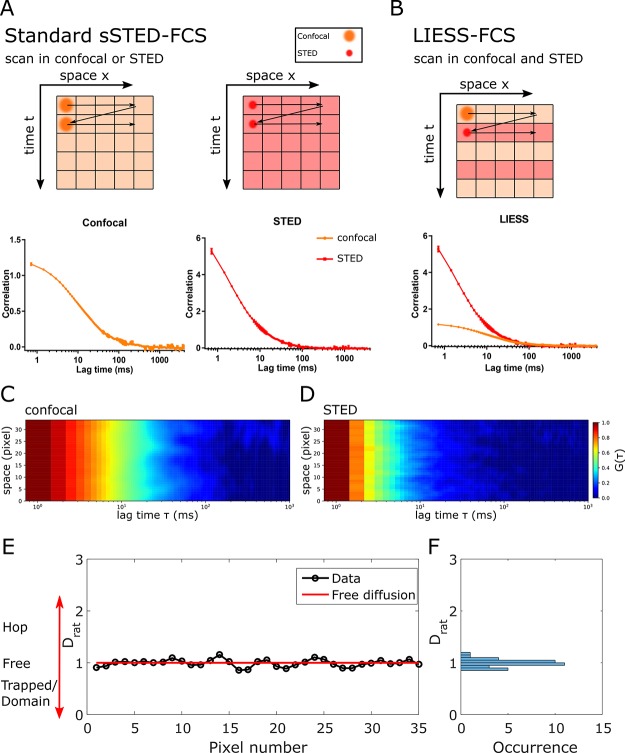
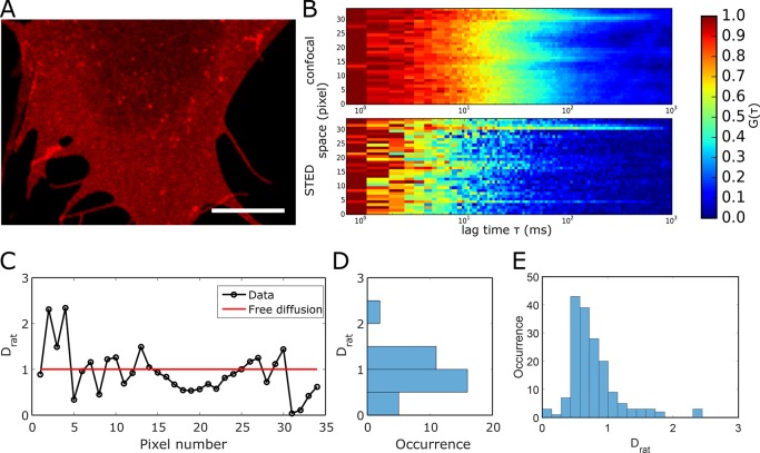
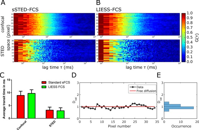
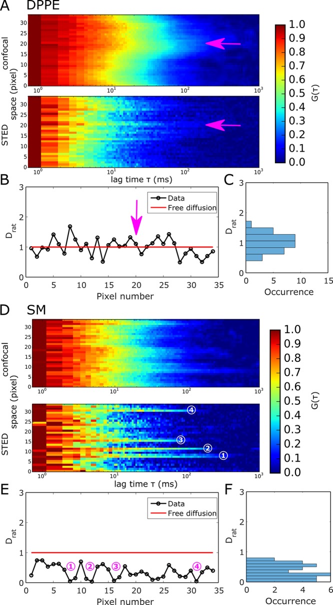
The diffusion dynamics in the cellular plasma membrane provide crucial insights into molecular interactions, organization, and bioactivity. Beam-scanning fluorescence correlation spectroscopy combined with super-resolution stimulated emission depletion nanoscopy (scanning STED-FCS) measures such dynamics with high spatial and temporal resolution. It reveals nanoscale diffusion characteristics by measuring the molecular diffusion in conventional confocal mode and super-resolved STED mode sequentially for each pixel along the scanned line. However, to directly link the spatial and the temporal information, a method that simultaneously measures the diffusion in confocal and STED modes is needed. Here, to overcome this problem, we establish an advanced STED-FCS measurement method, line interleaved excitation scanning STED-FCS (LIESS-FCS), that discloses the molecular diffusion modes at different spatial positions with a single measurement. It relies on fast beam-scanning along a line with alternating laser illumination that yields, for each pixel, the apparent diffusion coefficients for two different observation spot sizes (conventional confocal and super-resolved STED). We demonstrate the potential of the LIESS-FCS approach with simulations and experiments on lipid diffusion in model and live cell plasma membranes. We also apply LIESS-FCS to investigate the spatiotemporal organization of glycosylphosphatidylinositol-anchored proteins in the plasma membrane of live cells, which, interestingly, show multiple diffusion modes at different spatial positions.
细胞质膜中的扩散动力学为分子相互作用、组织和生物活性提供了重要的见解。基于扫描的荧光相关光谱学与超分辨率受激发射损耗纳米显微镜术(扫描 STED-FCS)相结合,可以以高时空分辨率测量此类动力学。它通过在传统共聚焦模式和超分辨 STED 模式下依次测量沿扫描线的每个像素的分子扩散,来揭示纳米级扩散特性。然而,为了直接关联空间和时间信息,需要一种能够同时测量共聚焦和 STED 模式下扩散的方法。在这里,为了克服这个问题,我们建立了一种先进的 STED-FCS 测量方法,即线交错激发扫描 STED-FCS(LIESS-FCS),它可以通过单次测量在不同的空间位置揭示分子扩散模式。它依赖于沿着一条线的快速光束扫描,交替激光照明,从而为每个像素提供两种不同观察光斑尺寸(传统共聚焦和超分辨 STED)的表观扩散系数。我们通过模拟和模型及活细胞膜中脂质扩散的实验,证明了 LIESS-FCS 方法的潜力。我们还将 LIESS-FCS 应用于研究活细胞质膜中糖基磷脂酰肌醇锚定蛋白的时空组织,有趣的是,它在不同的空间位置显示出多种扩散模式。