• 文献检索
  • 文档翻译
  • 深度研究
  • 学术资讯
  • Suppr Zotero 插件Zotero 插件
  • 邀请有礼
  • 套餐&价格
  • 历史记录
应用&插件
Suppr Zotero 插件Zotero 插件浏览器插件Mac 客户端Windows 客户端微信小程序
定价
高级版会员购买积分包购买API积分包
服务
文献检索文档翻译深度研究API 文档MCP 服务
关于我们
关于 Suppr公司介绍联系我们用户协议隐私条款
关注我们

Suppr 超能文献

核心技术专利:CN118964589B侵权必究
粤ICP备2023148730 号-1Suppr @ 2026

文献检索

告别复杂PubMed语法,用中文像聊天一样搜索,搜遍4000万医学文献。AI智能推荐,让科研检索更轻松。

立即免费搜索

文件翻译

保留排版,准确专业,支持PDF/Word/PPT等文件格式,支持 12+语言互译。

免费翻译文档

深度研究

AI帮你快速写综述,25分钟生成高质量综述,智能提取关键信息,辅助科研写作。

立即免费体验

量化梗死生长和二次损伤体积:比较多模态图像配准测量。

Quantifying Infarct Growth and Secondary Injury Volumes: Comparing Multimodal Image Registration Measures.

机构信息

From the Radcliffe Department of Medicine, Acute Vascular Imaging Centre (G.W.J.H., D.C., F.S., J.K.)

From the Radcliffe Department of Medicine, Acute Vascular Imaging Centre (G.W.J.H., D.C., F.S., J.K.).

出版信息

Stroke. 2018 Jul;49(7):1647-1655. doi: 10.1161/STROKEAHA.118.020788. Epub 2018 Jun 12.

DOI:10.1161/STROKEAHA.118.020788
PMID:29895538
原文链接:https://pmc.ncbi.nlm.nih.gov/articles/PMC6023577/
Abstract

BACKGROUND AND PURPOSE

Lesion expansion in the week after acute stroke involves both infarct growth (IG) and anatomic distortion (AD) because of edema and hemorrhage. Enabling separate quantification would allow clinical trials targeting these distinct pathological processes. We developed an objective and automated approach to quantify these processes at 24 hours and 1 week.

METHODS

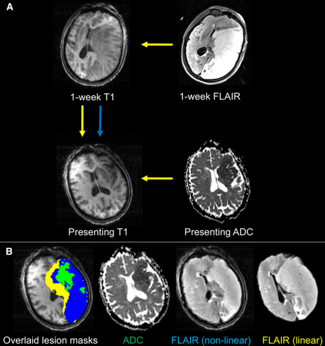
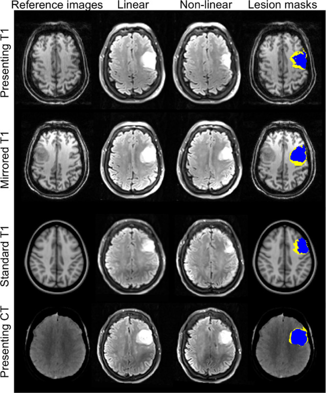
Patients with acute ischemic stroke were scanned at presentation, 24 hours, and 1 week in a magnetic resonance imaging (MRI) cohort study. IG and AD were calculated from follow-up lesion masks after linear and nonlinear registration to a presenting MRI scan. Performance of IG and AD was compared with edema quantified using cerebrospinal fluid displacement. The use of alternative reference images to define AD, including template MRI, mirrored MRI, and presenting computed tomographic scan, was explored.

RESULTS

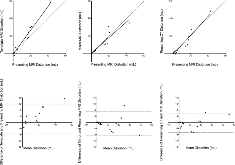
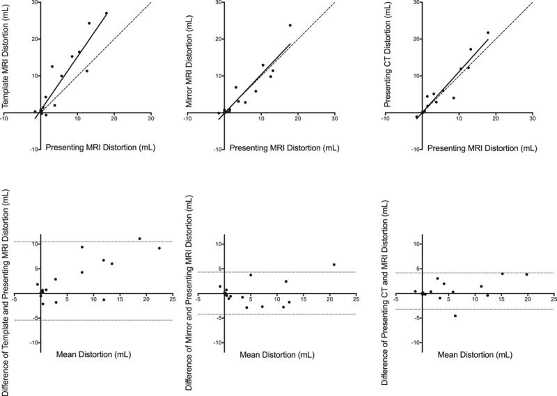
Thirty-seven patients with nonlacunar stroke were included. AD was responsible for 20% and 36% of lesion expansion at 24 hours (n=30) and 1 week (n=28). Registration-defined IG and AD compared favorably with edema quantified using cerebrospinal fluid displacement, particularly at smaller infarct volumes. Presenting computed tomographic imaging was the preferred alternative reference image to presenting MRI for measuring AD.

CONCLUSIONS

The contributions of IG and AD to lesion expansion can be measured separately over time through the use of image registration. This approach can be used to combine imaging outcome data from computed tomography and MRI.

摘要

背景与目的

急性卒中后一周内的病灶扩大包括梗死生长(IG)和由于水肿和出血导致的解剖变形(AD)。能够对这两种过程进行单独定量将允许临床试验针对这些不同的病理过程。我们开发了一种客观且自动化的方法,可在 24 小时和 1 周时定量测量这两种过程。

方法

在一项磁共振成像(MRI)队列研究中,对急性缺血性卒中患者在发病时、24 小时和 1 周时进行扫描。IG 和 AD 通过从后续病变掩模到初始 MRI 扫描的线性和非线性配准进行计算。将 IG 和 AD 的性能与使用脑脊液位移定量的水肿进行了比较。还探索了使用替代参考图像(包括模板 MRI、镜像 MRI 和初始 CT 扫描)来定义 AD。

结果

共纳入 37 例非腔隙性卒中患者。在 24 小时(n=30)和 1 周(n=28)时,AD 分别导致 20%和 36%的病灶扩大。通过图像配准定义的 IG 和 AD 与使用脑脊液位移定量的水肿具有良好的可比性,特别是在较小的梗死体积时。与初始 MRI 相比,初始 CT 成像作为 AD 的替代参考图像更为可取。

结论

通过使用图像配准,可以随时间分别测量 IG 和 AD 对病灶扩大的贡献。该方法可用于结合 CT 和 MRI 的成像结果数据。

https://cdn.ncbi.nlm.nih.gov/pmc/blobs/3861/6023577/3e56037bcd77/str-49-1647-g006.jpg
https://cdn.ncbi.nlm.nih.gov/pmc/blobs/3861/6023577/997a5b05d1a2/str-49-1647-g001.jpg
https://cdn.ncbi.nlm.nih.gov/pmc/blobs/3861/6023577/d7b1e86c6c4c/str-49-1647-g003.jpg
https://cdn.ncbi.nlm.nih.gov/pmc/blobs/3861/6023577/a9d56fc0196d/str-49-1647-g005.jpg
https://cdn.ncbi.nlm.nih.gov/pmc/blobs/3861/6023577/3e56037bcd77/str-49-1647-g006.jpg

相似文献

1
Quantifying Infarct Growth and Secondary Injury Volumes: Comparing Multimodal Image Registration Measures.量化梗死生长和二次损伤体积:比较多模态图像配准测量。
Stroke. 2018 Jul;49(7):1647-1655. doi: 10.1161/STROKEAHA.118.020788. Epub 2018 Jun 12.
2
Subacute Infarct Volume With Edema Correction in Computed Tomography Is Equivalent to Final Infarct Volume After Ischemic Stroke: Improving the Comparability of Infarct Imaging Endpoints in Clinical Trials.计算机断层扫描中的亚急性梗死体积与水肿校正相当于缺血性脑卒中后的最终梗死体积:提高临床试验中梗死成像终点的可比性。
Invest Radiol. 2018 Aug;53(8):472-476. doi: 10.1097/RLI.0000000000000475.
3
Quantitative Lesion Water Uptake in Acute Stroke Computed Tomography Is a Predictor of Malignant Infarction.急性脑卒中 CT 定量病变水摄取与恶性梗死相关。
Stroke. 2018 Aug;49(8):1906-1912. doi: 10.1161/STROKEAHA.118.020507.
4
Prediction of final infarct volume on subacute MRI by quantifying cerebral edema in ischemic stroke.通过量化缺血性卒中的脑水肿,利用亚急性磁共振成像预测最终梗死体积。
J Cereb Blood Flow Metab. 2017 Aug;37(8):3077-3084. doi: 10.1177/0271678X16683960. Epub 2016 Jan 1.
5
Comparison of perfusion computed tomography with diffusion-weighted magnetic resonance imaging in hyperacute ischemic stroke.超急性缺血性卒中中灌注计算机断层扫描与扩散加权磁共振成像的比较
J Comput Assist Tomogr. 2004 Nov-Dec;28(6):747-55. doi: 10.1097/00004728-200411000-00004.
6
Reduction in Cerebrospinal Fluid Volume as an Early Quantitative Biomarker of Cerebral Edema After Ischemic Stroke.脑血容量减少作为缺血性脑卒中后脑水肿的早期定量生物标志物。
Stroke. 2020 Feb;51(2):462-467. doi: 10.1161/STROKEAHA.119.027895. Epub 2019 Dec 10.
7
Early neurological stability predicts adverse outcome after acute ischemic stroke.早期神经学稳定性可预测急性缺血性卒中后的不良结局。
Int J Stroke. 2016 Oct;11(8):882-889. doi: 10.1177/1747493016654484. Epub 2016 Jul 9.
8
Unenhanced Dual-Energy Computed Tomography: Visualization of Brain Edema.未增强双能 CT 扫描:脑水肿的可视化。
Invest Radiol. 2018 Feb;53(2):63-69. doi: 10.1097/RLI.0000000000000413.
9
Ischemic lesion growth in acute stroke: Water uptake quantification distinguishes between edema and tissue infarct.急性卒中缺血性病变进展:水摄取定量可区分水肿和组织梗死。
J Cereb Blood Flow Metab. 2020 Apr;40(4):823-832. doi: 10.1177/0271678X19848505. Epub 2019 May 9.
10
"X-Map 2.0" for Edema Signal Enhancement for Acute Ischemic Stroke Using Non-Contrast-Enhanced Dual-Energy Computed Tomography.基于非对比增强双能量 CT 的急性缺血性脑卒中水肿信号增强的“X-Map 2.0”。
Invest Radiol. 2018 Jul;53(7):432-439. doi: 10.1097/RLI.0000000000000461.

引用本文的文献

1
Automatically quantified follow-up imaging biomarkers predict clinical outcomes after acute ischemic stroke.自动定量的随访成像生物标志物可预测急性缺血性中风后的临床结局。
Front Neurol. 2025 Mar 19;16:1483138. doi: 10.3389/fneur.2025.1483138. eCollection 2025.
2
Safety and efficacy of Angong Niuhuang Pills in patients with moderate-to-severe acute ischemic stroke (ANGONG TRIAL): A randomized double-blind placebo-controlled pilot clinical trial.安宫牛黄丸治疗中重度急性缺血性脑卒中患者的安全性和有效性(安宫试验):一项随机双盲安慰剂对照的前瞻性临床试验。
Chin Med J (Engl). 2024 Nov 6;138(5):579-88. doi: 10.1097/CM9.0000000000003133.
3

本文引用的文献

1
Thrombectomy 6 to 24 Hours after Stroke with a Mismatch between Deficit and Infarct.发病后 6 至 24 小时内进行取栓术治疗与缺损和梗死不匹配的脑卒中。
N Engl J Med. 2018 Jan 4;378(1):11-21. doi: 10.1056/NEJMoa1706442. Epub 2017 Nov 11.
2
Atlas registration for edema-corrected MRI lesion volume in mouse stroke models.针对小鼠中风模型的水肿校正 MRI 病变体积的图谱配准。
J Cereb Blood Flow Metab. 2019 Feb;39(2):313-323. doi: 10.1177/0271678X17726635. Epub 2017 Aug 22.
3
Optimizing image registration and infarct definition in stroke research.
Acute ischemic STROKE - from laboratory to the Patient's BED (STROKELABED): A translational approach to reperfusion injury. Study Protocol.
急性缺血性卒中——从实验室到患者病床(STROKELABED):再灌注损伤的转化研究方法。研究方案。
Transl Neurosci. 2024 Jul 12;15(1):20220344. doi: 10.1515/tnsci-2022-0344. eCollection 2024 Jan 1.
4
Late Lesion Growth following Endovascular Therapy: Is 24 h Too Early to Assess Acute Infarct Size Including the Effects of Secondary Injury?血管内治疗后晚期病变生长:评估急性梗死面积(包括继发性损伤的影响),24小时是否太早?
Cerebrovasc Dis. 2025;54(1):129-137. doi: 10.1159/000536470. Epub 2024 Feb 27.
5
Analysis of brain edema in RHAPSODY.RHAPSODY 中脑水肿的分析。
Int J Stroke. 2024 Jan;19(1):68-75. doi: 10.1177/17474930231187268. Epub 2023 Jul 16.
6
Infarct Growth despite Successful Endovascular Reperfusion in Acute Ischemic Stroke: A Meta-analysis.急性缺血性脑卒中血管内再通成功后仍出现梗死进展:一项荟萃分析。
AJNR Am J Neuroradiol. 2021 Aug;42(8):1472-1478. doi: 10.3174/ajnr.A7177. Epub 2021 Jun 3.
7
Association between pre-treatment perfusion profile and cerebral edema after reperfusion therapies in ischemic stroke.缺血性脑卒中再灌注治疗后预处理灌注谱与脑水肿的关系。
J Cereb Blood Flow Metab. 2021 Nov;41(11):2887-2896. doi: 10.1177/0271678X211017696. Epub 2021 May 17.
8
PET-MRI nanoparticles imaging of blood-brain barrier damage and modulation after stroke reperfusion.正电子发射断层扫描-磁共振成像纳米颗粒对中风再灌注后脑血屏障损伤及调节的成像
Brain Commun. 2020 Nov 11;2(2):fcaa193. doi: 10.1093/braincomms/fcaa193. eCollection 2020.
9
Matrix Metalloproteinase-9 Relationship With Infarct Growth and Hemorrhagic Transformation in the Era of Thrombectomy.血栓切除术时代基质金属蛋白酶-9与梗死灶扩大及出血性转化的关系
Front Neurol. 2020 Jun 9;11:473. doi: 10.3389/fneur.2020.00473. eCollection 2020.
10
A non-human primate model of stroke reproducing endovascular thrombectomy and allowing long-term imaging and neurological read-outs.一种模拟血管内血栓切除术的非人类灵长类中风模型,可进行长期影像学和神经学检测。
J Cereb Blood Flow Metab. 2021 Apr;41(4):745-760. doi: 10.1177/0271678X20921310. Epub 2020 May 19.
优化中风研究中的图像配准和梗死灶定义。
Ann Clin Transl Neurol. 2017 Jan 20;4(3):166-174. doi: 10.1002/acn3.388. eCollection 2017 Mar.
4
Prediction of final infarct volume on subacute MRI by quantifying cerebral edema in ischemic stroke.通过量化缺血性卒中的脑水肿,利用亚急性磁共振成像预测最终梗死体积。
J Cereb Blood Flow Metab. 2017 Aug;37(8):3077-3084. doi: 10.1177/0271678X16683960. Epub 2016 Jan 1.
5
Safety and efficacy of intravenous glyburide on brain swelling after large hemispheric infarction (GAMES-RP): a randomised, double-blind, placebo-controlled phase 2 trial.大型半球性脑梗死患者静脉用格列本脲减轻脑水肿的安全性和有效性研究(GAMES-RP):一项随机、双盲、安慰剂对照的 2 期临床试验。
Lancet Neurol. 2016 Oct;15(11):1160-9. doi: 10.1016/S1474-4422(16)30196-X. Epub 2016 Aug 23.
6
Comparison between voxel-based and subtraction methods for measuring diffusion-weighted imaging lesion growth after thrombolysis.基于体素与减影方法测量溶栓后弥散加权成像病灶增长的比较。
Int J Stroke. 2016 Feb;11(2):221-8. doi: 10.1177/1747493015616636.
7
Novel Imaging Markers of Ischemic Cerebral Edema and Its Association with Neurological Outcome.缺血性脑水肿的新型成像标志物及其与神经功能结局的关联
Acta Neurochir Suppl. 2016;121:223-6. doi: 10.1007/978-3-319-18497-5_40.
8
Challenges and Opportunities of Endovascular Stroke Therapy.血管内卒中治疗的挑战与机遇。
Ann Neurol. 2016 Jan;79(1):11-7. doi: 10.1002/ana.24528. Epub 2015 Dec 15.
9
Stent-retriever thrombectomy after intravenous t-PA vs. t-PA alone in stroke.血管内溶栓联合支架取栓与单纯静脉溶栓治疗脑卒中的比较。
N Engl J Med. 2015 Jun 11;372(24):2285-95. doi: 10.1056/NEJMoa1415061. Epub 2015 Apr 17.
10
Know your tools--concordance of different methods for measuring brain volume change after ischemic stroke.了解你的工具——缺血性中风后测量脑容量变化的不同方法的一致性
Neuroradiology. 2015 Jul;57(7):685-95. doi: 10.1007/s00234-015-1522-8. Epub 2015 Apr 8.