Debatisse Justine, Eker Omer Faruk, Wateau Océane, Cho Tae-Hee, Wiart Marlène, Ramonet David, Costes Nicolas, Mérida Inés, Léon Christelle, Dia Maya, Paillard Mélanie, Confais Joachim, Rossetti Fabien, Langlois Jean-Baptiste, Troalen Thomas, Iecker Thibaut, Le Bars Didier, Lancelot Sophie, Bouchier Baptiste, Lukasziewicz Anne-Claire, Oudotte Adrien, Nighoghossian Norbert, Ovize Michel, Contamin Hugues, Lux François, Tillement Olivier, Canet-Soulas Emmanuelle
Univ Lyon, CarMeN Laboratory, INSERM, INRA, INSA Lyon, Université Claude Bernard Lyon 1, 69000 Lyon, France.
Siemens-Healthcare SAS, Saint-Denis, France.
Brain Commun. 2020 Nov 11;2(2):fcaa193. doi: 10.1093/braincomms/fcaa193. eCollection 2020.
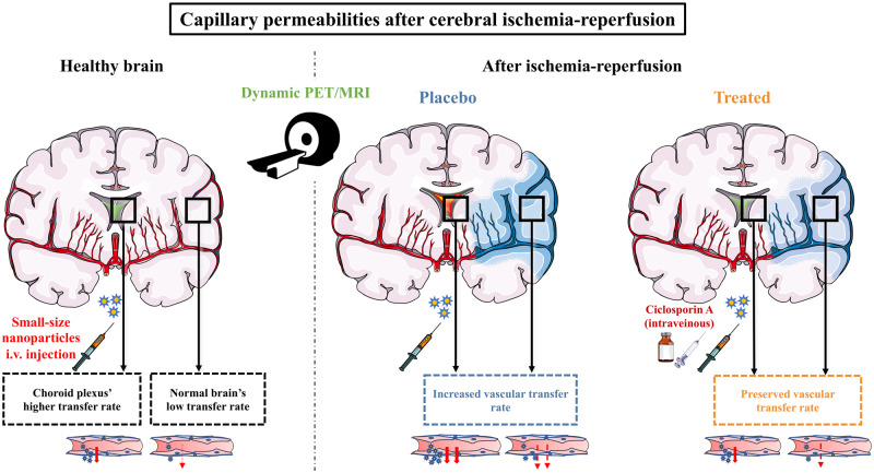
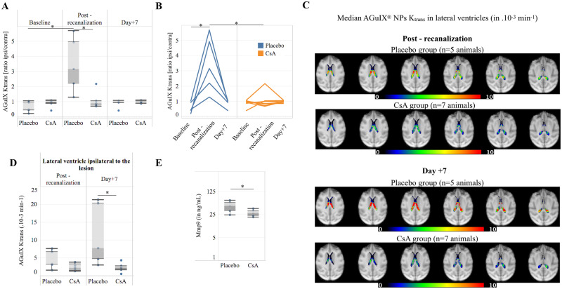
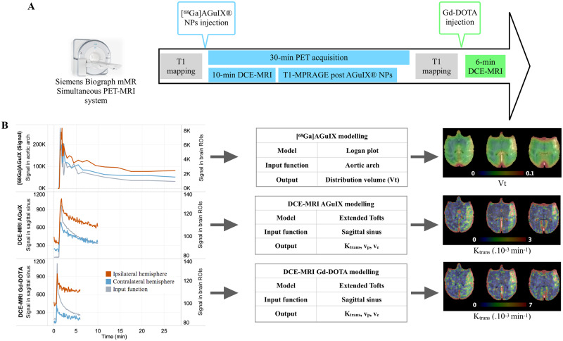
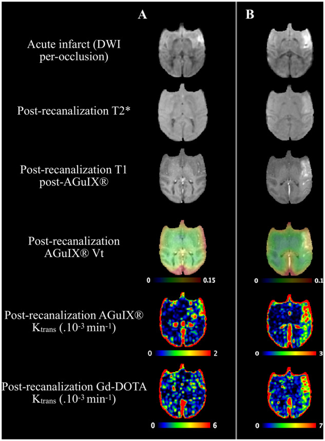
In an acute ischaemic stroke, understanding the dynamics of blood-brain barrier injury is of particular importance for the prevention of symptomatic haemorrhagic transformation. However, the available techniques assessing blood-brain barrier permeability are not quantitative and are little used in the context of acute reperfusion therapy. Nanoparticles cross the healthy or impaired blood-brain barrier through combined passive and active processes. Imaging and quantifying their transfer rate could better characterize blood-brain barrier damage and refine the delivery of neuroprotective agents. We previously developed an original endovascular stroke model of acute ischaemic stroke treated by mechanical thrombectomy followed by positron emission tomography-magnetic resonance imaging. Cerebral capillary permeability was quantified for two molecule sizes: small clinical gadolinium Gd-DOTA (<1 nm) and AGuIX nanoparticles (∼5 nm) used for brain theranostics. On dynamic contrast-enhanced magnetic resonance imaging, the baseline transfer constant was 0.94 [0.48, 1.72] and 0.16 [0.08, 0.33] ×10min, respectively, in the normal brain parenchyma, consistent with their respective sizes, and 1.90 [1.23, 3.95] and 2.86 [1.39, 4.52] ×10min in choroid plexus, confirming higher permeability than brain parenchyma. At early reperfusion, for both Gd-DOTA and AGuIX nanoparticles was significantly higher within the ischaemic area compared to the contralateral hemisphere; 2.23 [1.17, 4.13] and 0.82 [0.46, 1.87] ×10min for Gd-DOTA and AGuIX nanoparticles, respectively. With AGuIX nanoparticles, also increased within the ischaemic growth areas, suggesting added value for AGuIX. Finally, was significantly lower in both the lesion and the choroid plexus in a drug-treated group (ciclosporin A, = 7) compared to placebo ( = 5). quantification with AGuIX nanoparticles can monitor early blood-brain barrier damage and treatment effect in ischaemic stroke after reperfusion.
在急性缺血性卒中中,了解血脑屏障损伤的动态变化对于预防症状性出血转化尤为重要。然而,现有的评估血脑屏障通透性的技术并非定量的,且在急性再灌注治疗的背景下很少使用。纳米颗粒通过被动和主动相结合的过程穿过健康或受损的血脑屏障。对其转运速率进行成像和定量分析可以更好地表征血脑屏障损伤,并优化神经保护剂的递送。我们之前开发了一种原创的急性缺血性卒中血管内模型,通过机械取栓术治疗,随后进行正电子发射断层扫描-磁共振成像。针对两种分子大小对脑毛细血管通透性进行了量化:临床常用的小分子钆喷酸葡胺(Gd-DOTA,<1 nm)和用于脑诊疗的AGuIX纳米颗粒(~5 nm)。在动态对比增强磁共振成像中,正常脑实质内Gd-DOTA和AGuIX纳米颗粒的基线转运常数分别为0.94 [0.48, 1.72] 和0.16 [0.08, 0.33]×10⁻³ min⁻¹,与其各自大小相符,而脉络丛中的转运常数分别为1.90 [1.23, 3.95] 和2.86 [1.39, 4.52]×10⁻³ min⁻¹,证实脉络丛的通透性高于脑实质。在早期再灌注时,缺血区域内Gd-DOTA和AGuIX纳米颗粒的转运常数均显著高于对侧半球;Gd-DOTA和AGuIX纳米颗粒的转运常数分别为2.23 [1.17, 4.13] 和0.82 [0.46, 1.87]×10⁻³ min⁻¹。对于AGuIX纳米颗粒,缺血扩展区域内的转运常数也有所增加,表明AGuIX具有额外价值。最后,与安慰剂组(n = 5)相比,药物治疗组(环孢素A,n = 7)的病变区域和脉络丛中的转运常数均显著降低。使用AGuIX纳米颗粒进行转运常数量化可以监测再灌注后缺血性卒中早期的血脑屏障损伤和治疗效果。