Department of Oral and Maxillofacial Pathology, School of Dentistry, Kyung Hee University, Seoul 02447, Korea.
Department of Dentistry, Graduate School, Kyung Hee University, Seoul 02447, Korea.
Int J Mol Sci. 2018 Jun 12;19(6):1742. doi: 10.3390/ijms19061742.
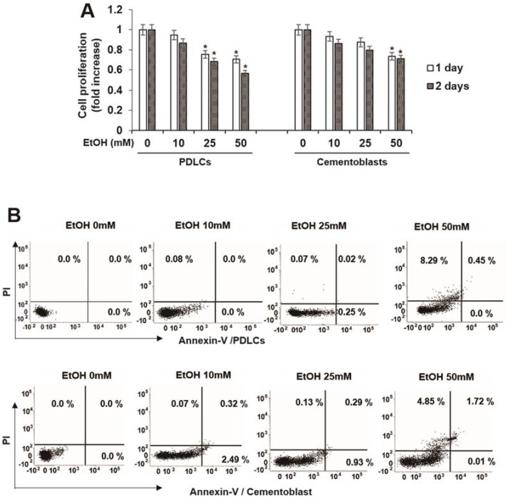
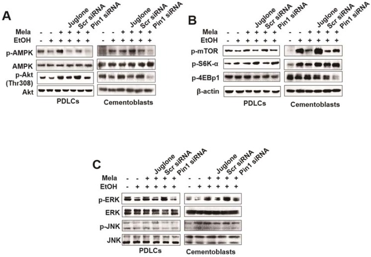
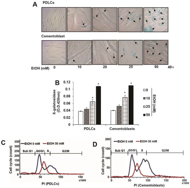
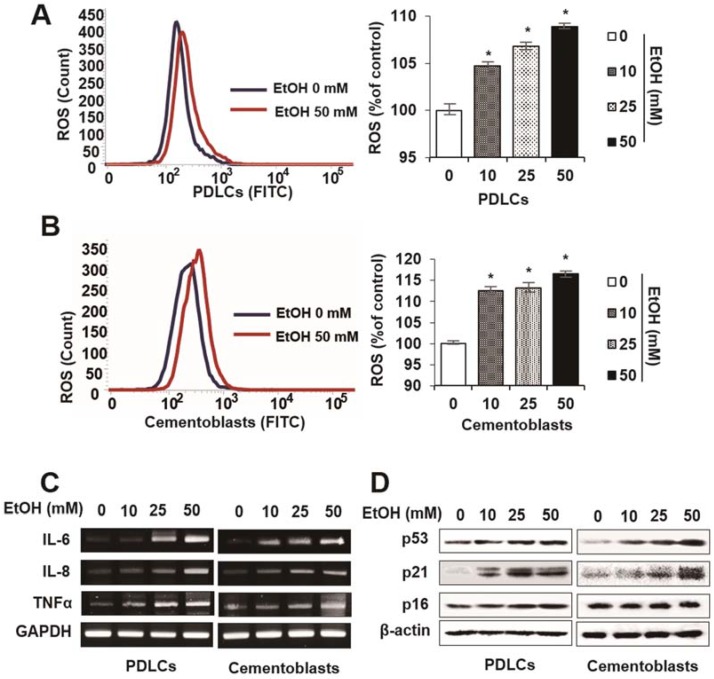
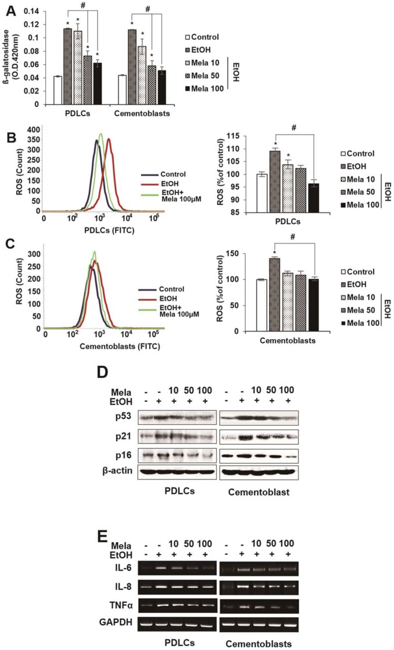
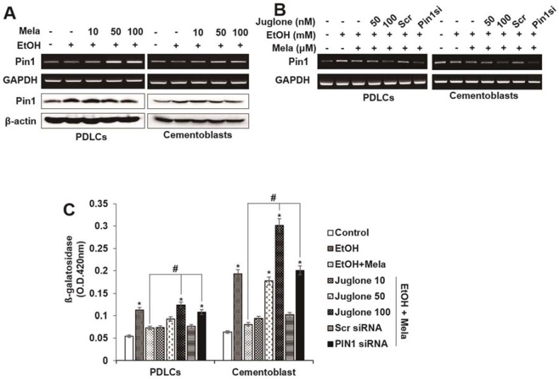
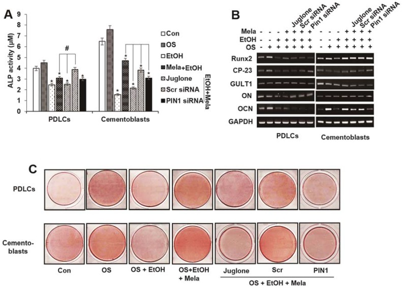
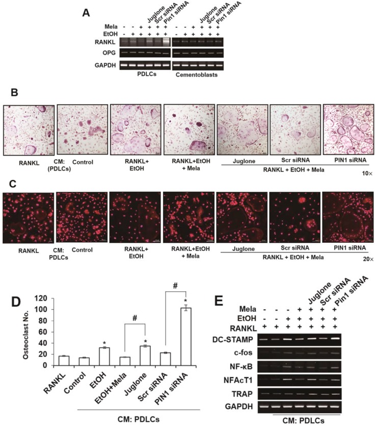
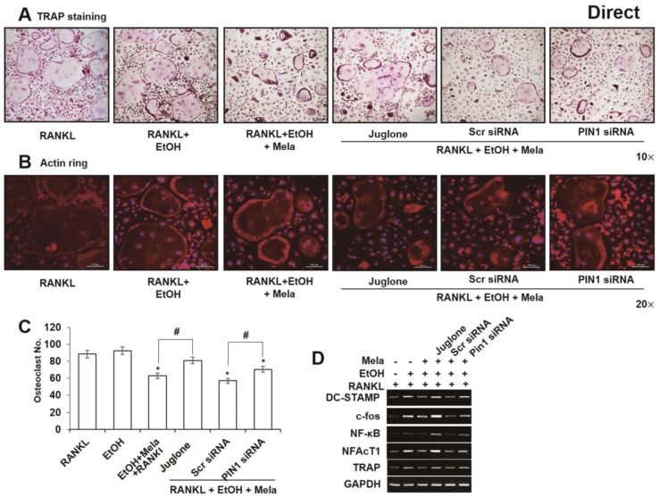
The present study evaluated the protective effects of melatonin in ethanol (EtOH)-induced senescence and osteoclastic differentiation in human periodontal ligament cells (HPDLCs) and cementoblasts and the underlying mechanism. EtOH increased senescence activity, levels of reactive oxygen species (ROS) and the expression of cell cycle regulators (p53, p21 and p16) and senescence-associated secretory phenotype () genes (interleukin [IL]-1β, IL-6, IL-8 and tumor necrosis factor-α) in HPDLCs and cementoblasts. Melatonin inhibited EtOH-induced senescence and the production of ROS as well as the increased expression of cell cycle regulators and SASP genes. However, it recovered EtOH-suppressed osteoblastic/cementoblastic differentiation, as evidenced by alkaline phosphatase activity, alizarin staining and mRNA expression levels of Runt-related transcription factor 2 (Runx2) and osteoblastic and cementoblastic markers (glucose transporter 1 and cementum-derived protein-32) in HPDLCs and cementoblasts. Moreover, it inhibited EtOH-induced osteoclastic differentiation in mouse bone marrow⁻derived macrophages (BMMs). Inhibition of protein never in mitosis gene A interacting-1 (PIN1) by juglone or small interfering RNA reversed the effects of melatonin on EtOH-mediated senescence as well as osteoblastic and osteoclastic differentiation. Melatonin blocked EtOH-induced activation of mammalian target of rapamycin (mTOR), AMP-activated protein kinase (AMPK), mitogen-activated protein kinase (MAPK) and Nuclear factor of activated T-cells (NFAT) c-1 pathways, which was reversed by inhibition of PIN1. This is the first study to show the protective effects of melatonin on senescence-like phenotypes and osteoclastic differentiation induced by oxidative stress in HPDLCs and cementoblasts through the PIN1 pathway.
本研究评估了褪黑素对人牙周韧带细胞(HPDLCs)和成牙骨质细胞中乙醇(EtOH)诱导的衰老和破骨细胞分化的保护作用及其机制。乙醇增加了 HPDLCs 和成牙骨质细胞的衰老活性、活性氧(ROS)水平以及细胞周期调节剂(p53、p21 和 p16)和衰老相关分泌表型(SASP)基因(白细胞介素[IL]-1β、IL-6、IL-8 和肿瘤坏死因子-α)的表达。褪黑素抑制 EtOH 诱导的衰老和 ROS 的产生,以及细胞周期调节剂和 SASP 基因的表达增加。然而,它恢复了 EtOH 抑制的成骨/成牙骨质分化,这表现在 HPDLCs 和成牙骨质细胞中碱性磷酸酶活性、茜素红染色和 Runt 相关转录因子 2(Runx2)和成骨细胞和成牙骨质细胞标志物(葡萄糖转运蛋白 1 和牙骨质蛋白-32)的 mRNA 表达水平上。此外,它抑制了 EtOH 诱导的小鼠骨髓源性巨噬细胞(BMMs)中的破骨细胞分化。通过 Juglone 或小干扰 RNA 抑制蛋白激酶 C-N 端结构域相互作用蛋白 1(PIN1)逆转了褪黑素对 EtOH 介导的衰老以及成骨细胞和破骨细胞分化的影响。褪黑素阻断了 EtOH 诱导的哺乳动物雷帕霉素靶蛋白(mTOR)、AMP 激活的蛋白激酶(AMPK)、丝裂原激活的蛋白激酶(MAPK)和激活 T 细胞的核因子(NFAT)c-1 途径的激活,而 PIN1 的抑制则逆转了这一过程。这是第一项研究表明,褪黑素通过 PIN1 途径对 HPDLCs 和成牙骨质细胞中由氧化应激引起的衰老样表型和破骨细胞分化具有保护作用。