• 文献检索
  • 文档翻译
  • 深度研究
  • 学术资讯
  • Suppr Zotero 插件Zotero 插件
  • 邀请有礼
  • 套餐&价格
  • 历史记录
应用&插件
Suppr Zotero 插件Zotero 插件浏览器插件Mac 客户端Windows 客户端微信小程序
定价
高级版会员购买积分包购买API积分包
服务
文献检索文档翻译深度研究API 文档MCP 服务
关于我们
关于 Suppr公司介绍联系我们用户协议隐私条款
关注我们

Suppr 超能文献

核心技术专利:CN118964589B侵权必究
粤ICP备2023148730 号-1Suppr @ 2026

文献检索

告别复杂PubMed语法,用中文像聊天一样搜索,搜遍4000万医学文献。AI智能推荐,让科研检索更轻松。

立即免费搜索

文件翻译

保留排版,准确专业,支持PDF/Word/PPT等文件格式,支持 12+语言互译。

免费翻译文档

深度研究

AI帮你快速写综述,25分钟生成高质量综述,智能提取关键信息,辅助科研写作。

立即免费体验

青光眼患者视觉通路及脑连接的变化:一项系统评价

Changes of Visual Pathway and Brain Connectivity in Glaucoma: A Systematic Review.

作者信息

Nuzzi Raffaele, Dallorto Laura, Rolle Teresa

机构信息

Eye Clinic, Department of Surgical Sciences, University of Torino, Turin, Italy.

出版信息

Front Neurosci. 2018 May 29;12:363. doi: 10.3389/fnins.2018.00363. eCollection 2018.

DOI:10.3389/fnins.2018.00363
PMID:29896087
原文链接:https://pmc.ncbi.nlm.nih.gov/articles/PMC5986964/
Abstract

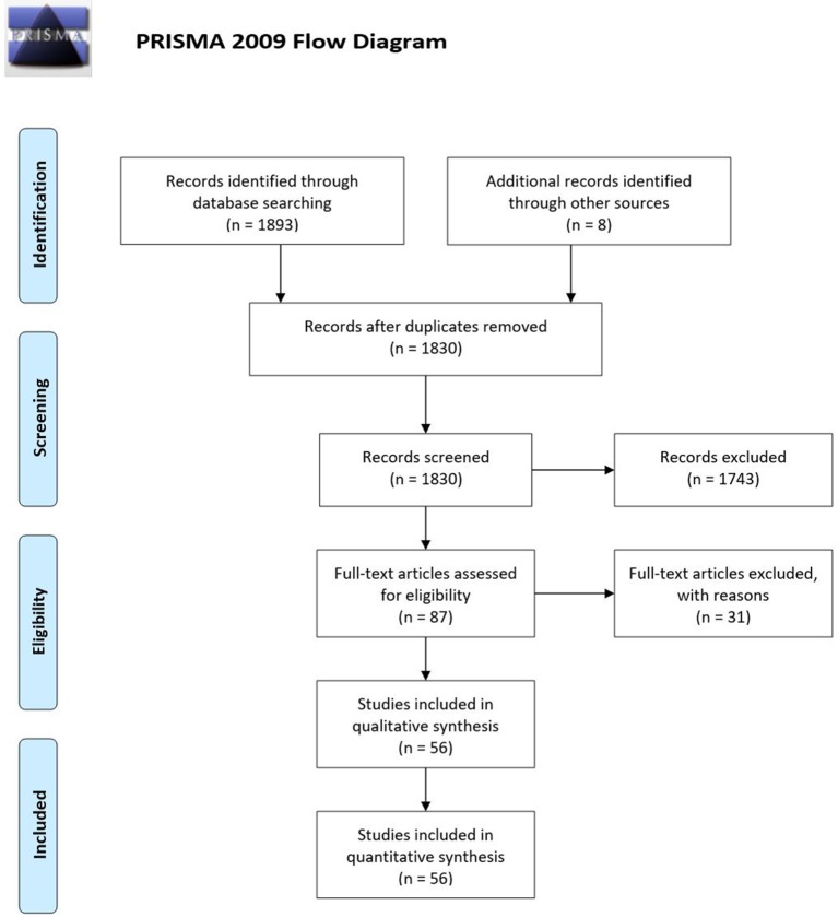
Glaucoma is a leading cause of irreversible blindness worldwide. The increasing interest in the involvement of the cortical visual pathway in glaucomatous patients is due to the implications in recent therapies, such as neuroprotection and neuroregeneration. In this review, we outline the current understanding of brain structural, functional, and metabolic changes detected with the modern techniques of neuroimaging in glaucomatous subjects. We screened MEDLINE, EMBASE, CINAHL, CENTRAL, LILACS, Trip Database, and NICE for original contributions published until 31 October 2017. Studies with at least six patients affected by any type of glaucoma were considered. We included studies using the following neuroimaging techniques: functional Magnetic Resonance Imaging (fMRI), resting-state fMRI (rs-fMRI), magnetic resonance spectroscopy (MRS), voxel- based Morphometry (VBM), surface-based Morphometry (SBM), diffusion tensor MRI (DTI). Over a total of 1,901 studies, 56 case series with a total of 2,381 patients were included. Evidence of neurodegenerative process in glaucomatous patients was found both within and beyond the visual system. Structural alterations in visual cortex (mainly reduced cortex thickness and volume) have been demonstrated with SBM and VBM; these changes were not limited to primary visual cortex but also involved association visual areas. Other brain regions, associated with visual function, demonstrated a certain grade of increased or decreased gray matter volume. Functional and metabolic abnormalities resulted within primary visual cortex in all studies with fMRI and MRS. Studies with rs-fMRI found disrupted connectivity between the primary and higher visual cortex and between visual cortex and associative visual areas in the task-free state of glaucomatous patients. This review contributes to the better understanding of brain abnormalities in glaucoma. It may stimulate further speculation about brain plasticity at a later age and therapeutic strategies, such as the prevention of cortical degeneration in patients with glaucoma. Structural, functional, and metabolic neuroimaging methods provided evidence of changes throughout the visual pathway in glaucomatous patients. Other brain areas, not directly involved in the processing of visual information, also showed alterations.

摘要

青光眼是全球不可逆性失明的主要原因。人们对青光眼患者皮质视觉通路受累的兴趣日益增加,这是由于其对近期治疗方法(如神经保护和神经再生)具有重要意义。在本综述中,我们概述了目前对青光眼患者使用现代神经影像学技术检测到的脑结构、功能和代谢变化的理解。我们在MEDLINE、EMBASE、CINAHL、CENTRAL、LILACS、Trip数据库和NICE中筛选了截至2017年10月31日发表的原创性研究。纳入了至少有6例受任何类型青光眼影响患者的研究。我们纳入了使用以下神经影像学技术的研究:功能磁共振成像(fMRI)、静息态fMRI(rs-fMRI)、磁共振波谱(MRS)、基于体素的形态学测量(VBM)、基于表面的形态学测量(SBM)、扩散张量MRI(DTI)。在总共1901项研究中,纳入了56个病例系列,共计2381例患者。在青光眼患者的视觉系统内外均发现了神经退行性变过程的证据。通过SBM和VBM已证实视觉皮层存在结构改变(主要是皮层厚度和体积减小);这些变化不仅限于初级视觉皮层,还涉及联合视觉区域。与视觉功能相关的其他脑区显示出一定程度的灰质体积增加或减少。在所有使用fMRI和MRS的研究中,初级视觉皮层内均出现了功能和代谢异常。rs-fMRI研究发现,在青光眼患者的无任务状态下,初级视觉皮层与高级视觉皮层之间以及视觉皮层与联合视觉区域之间的连接中断。本综述有助于更好地理解青光眼患者的脑异常情况。它可能会激发对晚年脑可塑性以及治疗策略(如预防青光眼患者的皮质变性)的进一步思考。结构、功能和代谢神经影像学方法为青光眼患者整个视觉通路的变化提供了证据。其他未直接参与视觉信息处理的脑区也显示出改变。

https://cdn.ncbi.nlm.nih.gov/pmc/blobs/e814/5986964/3e2e8762e914/fnins-12-00363-g0001.jpg

相似文献

1
Changes of Visual Pathway and Brain Connectivity in Glaucoma: A Systematic Review.青光眼患者视觉通路及脑连接的变化:一项系统评价
Front Neurosci. 2018 May 29;12:363. doi: 10.3389/fnins.2018.00363. eCollection 2018.
2
Reduced Functional and Anatomic Interhemispheric Homotopic Connectivity in Primary Open-Angle Glaucoma: A Combined Resting State-fMRI and DTI Study.原发性开角型青光眼患者功能和解剖性大脑半球间同型连接减少:一项静息态 fMRI 和 DTI 的联合研究。
Invest Ophthalmol Vis Sci. 2018 Apr 1;59(5):1861-1868. doi: 10.1167/iovs.17-23291.
3
Early changes of brain connectivity in primary open angle glaucoma.原发性开角型青光眼患者脑连接的早期变化
Hum Brain Mapp. 2016 Dec;37(12):4581-4596. doi: 10.1002/hbm.23330. Epub 2016 Aug 9.
4
[Aiming for zero blindness].追求零失明
Nippon Ganka Gakkai Zasshi. 2015 Mar;119(3):168-93; discussion 194.
5
Diffuse brain damage in normal tension glaucoma.正常眼压性青光眼中的弥漫性脑损伤。
Hum Brain Mapp. 2018 Jan;39(1):532-541. doi: 10.1002/hbm.23862. Epub 2017 Oct 24.
6
Primary Open Angle Glaucoma Is Associated With Functional Brain Network Reorganization.原发性开角型青光眼与功能性脑网络重组有关。
Front Neurol. 2019 Oct 25;10:1134. doi: 10.3389/fneur.2019.01134. eCollection 2019.
7
Magnetic resonance in studies of glaucoma.磁共振在青光眼研究中的应用。
Med Sci Monit. 2011 Oct;17(10):RA227-32. doi: 10.12659/msm.881973.
8
Central sensitization in fibromyalgia? A systematic review on structural and functional brain MRI.纤维肌痛中的中枢敏化?关于大脑结构和功能磁共振成像的系统综述
Semin Arthritis Rheum. 2014 Aug;44(1):68-75. doi: 10.1016/j.semarthrit.2014.01.001. Epub 2014 Jan 8.
9
Advanced Morphological and Functional Magnetic Resonance Techniques in Glaucoma.青光眼的先进形态学和功能磁共振技术
Biomed Res Int. 2015;2015:160454. doi: 10.1155/2015/160454. Epub 2015 Jun 8.
10
Glaucoma and the brain: Trans-synaptic degeneration, structural change, and implications for neuroprotection.青光眼与大脑:突触间变性、结构改变及其对神经保护的影响。
Surv Ophthalmol. 2018 May-Jun;63(3):296-306. doi: 10.1016/j.survophthal.2017.09.010. Epub 2017 Oct 3.

引用本文的文献

1
The Connectivity of the Resting Brain in Primary Open-Angle Glaucoma: A Systematic Review.原发性开角型青光眼静息态脑功能连接性的系统评价
Biomedicines. 2025 Jun 7;13(6):1402. doi: 10.3390/biomedicines13061402.
2
Microglia remodeling in the visual thalamus of the DBA/2J mouse model of glaucoma.青光眼DBA/2J小鼠模型视丘中小胶质细胞的重塑
PLoS One. 2025 May 15;20(5):e0323513. doi: 10.1371/journal.pone.0323513. eCollection 2025.
3
Glaucoma and brain functional networks: a bidirectional Mendelian randomisation study.青光眼与脑功能网络:一项双向孟德尔随机化研究

本文引用的文献

1
Diffuse brain damage in normal tension glaucoma.正常眼压性青光眼中的弥漫性脑损伤。
Hum Brain Mapp. 2018 Jan;39(1):532-541. doi: 10.1002/hbm.23862. Epub 2017 Oct 24.
2
Primary angle-closure glaucomas disturb regional spontaneous brain activity in the visual pathway: an fMRI study.原发性闭角型青光眼扰乱视觉通路中的局部自发性脑活动:一项功能磁共振成像研究。
Neuropsychiatr Dis Treat. 2017 May 25;13:1409-1417. doi: 10.2147/NDT.S134258. eCollection 2017.
3
Management of glaucoma as a neurodegenerative disease.作为神经退行性疾病的青光眼管理
BMJ Open Ophthalmol. 2025 Apr 12;10(1):e001902. doi: 10.1136/bmjophth-2024-001902.
4
Contrast Discrimination and Global Form Perception in Primary Open-Angle Glaucoma and Primary Angle-Closure Glaucoma.原发性开角型青光眼和原发性闭角型青光眼的对比辨色力和全局形态感知。
Invest Ophthalmol Vis Sci. 2024 May 1;65(5):33. doi: 10.1167/iovs.65.5.33.
5
Convolutional neural network-based classification of glaucoma using optic radiation tissue properties.基于卷积神经网络利用视辐射组织特性对青光眼进行分类
Commun Med (Lond). 2024 Apr 11;4(1):72. doi: 10.1038/s43856-024-00496-w.
6
Extraction of three-dimensional shapes in glaucoma patients in response to monocular depth cues.青光眼患者对单眼深度线索的三维形状提取。
Jpn J Ophthalmol. 2024 May;68(3):183-191. doi: 10.1007/s10384-024-01054-6. Epub 2024 Apr 10.
7
GDNet-EEG: An attention-aware deep neural network based on group depth-wise convolution for SSVEP stimulation frequency recognition.GDNet-EEG:一种基于分组深度卷积的注意力感知深度神经网络,用于稳态视觉诱发电位刺激频率识别。
Front Neurosci. 2023 Apr 13;17:1160040. doi: 10.3389/fnins.2023.1160040. eCollection 2023.
8
Magnetic resonance imaging assessment of the lateral geniculate nucleus volume and height in patients with glaucoma: a Meta-analysis.青光眼患者外侧膝状体体积和高度的磁共振成像评估:一项Meta分析
Int J Ophthalmol. 2023 Apr 18;16(4):638-644. doi: 10.18240/ijo.2023.04.19. eCollection 2023.
9
Altered dynamic functional connectivity in the primary visual cortex in patients with primary angle-closure glaucoma.原发性闭角型青光眼患者初级视觉皮层动态功能连接的改变
Front Neurosci. 2023 Feb 2;17:1131247. doi: 10.3389/fnins.2023.1131247. eCollection 2023.
10
High sampling resolution optical coherence tomography reveals potential concurrent reductions in ganglion cell-inner plexiform and inner nuclear layer thickness but not in outer retinal thickness in glaucoma.高采样分辨率光学相干断层扫描显示青光眼患者神经节细胞-内丛状层和内核层厚度可能同时减少,但外视网膜厚度无变化。
Ophthalmic Physiol Opt. 2023 Jan;43(1):46-63. doi: 10.1111/opo.13065. Epub 2022 Nov 23.
Neurodegener Dis Manag. 2017 Apr;7(2):157-172. doi: 10.2217/nmt-2017-0004. Epub 2017 May 22.
4
Experience-Dependent Structural Plasticity in the Visual System.视觉系统中的经验依赖性结构可塑性。
Annu Rev Vis Sci. 2016 Oct 14;2:17-35. doi: 10.1146/annurev-vision-111815-114638.
5
Resting state fMRI: A review on methods in resting state connectivity analysis and resting state networks.静息态功能磁共振成像:静息态连接性分析及静息态网络方法综述
Neuroradiol J. 2017 Aug;30(4):305-317. doi: 10.1177/1971400917697342. Epub 2017 Mar 29.
6
Structural and functional brain changes in early- and mid-stage primary open-angle glaucoma using voxel-based morphometry and functional magnetic resonance imaging.使用基于体素的形态测量法和功能磁共振成像技术对早中期原发性开角型青光眼患者脑结构和功能变化的研究
Medicine (Baltimore). 2017 Mar;96(9):e6139. doi: 10.1097/MD.0000000000006139.
7
Retinotopic fMRI Reveals Visual Dysfunction and Functional Reorganization in the Visual Cortex of Mild to Moderate Glaucoma Patients.视网膜拓扑功能磁共振成像揭示轻至中度青光眼患者视觉皮层的视觉功能障碍和功能重组。
J Glaucoma. 2017 May;26(5):430-437. doi: 10.1097/IJG.0000000000000641.
8
Neuroprotection for treatment of glaucoma in adults.用于治疗成人青光眼的神经保护。
Cochrane Database Syst Rev. 2017 Jan 25;1(1):CD006539. doi: 10.1002/14651858.CD006539.pub4.
9
Intrinsic Functional Connectivity Alterations of the Primary Visual Cortex in Primary Angle-Closure Glaucoma Patients before and after Surgery: A Resting-State fMRI Study.原发性闭角型青光眼患者手术前后初级视觉皮层的内在功能连接改变:一项静息态功能磁共振成像研究
PLoS One. 2017 Jan 25;12(1):e0170598. doi: 10.1371/journal.pone.0170598. eCollection 2017.
10
Optic Radiations Microstructural Changes in Glaucoma and Association With Severity: A Study Using 3Tesla-Magnetic Resonance Diffusion Tensor Imaging.青光眼视辐射的微观结构变化及其与严重程度的关联:一项使用3特斯拉磁共振扩散张量成像的研究
Invest Ophthalmol Vis Sci. 2016 Dec 1;57(15):6539-6547. doi: 10.1167/iovs.16-19838.