• 文献检索
  • 文档翻译
  • 深度研究
  • 学术资讯
  • Suppr Zotero 插件Zotero 插件
  • 邀请有礼
  • 套餐&价格
  • 历史记录
应用&插件
Suppr Zotero 插件Zotero 插件浏览器插件Mac 客户端Windows 客户端微信小程序
定价
高级版会员购买积分包购买API积分包
服务
文献检索文档翻译深度研究API 文档MCP 服务
关于我们
关于 Suppr公司介绍联系我们用户协议隐私条款
关注我们

Suppr 超能文献

核心技术专利:CN118964589B侵权必究
粤ICP备2023148730 号-1Suppr @ 2026

文献检索

告别复杂PubMed语法,用中文像聊天一样搜索,搜遍4000万医学文献。AI智能推荐,让科研检索更轻松。

立即免费搜索

文件翻译

保留排版,准确专业,支持PDF/Word/PPT等文件格式,支持 12+语言互译。

免费翻译文档

深度研究

AI帮你快速写综述,25分钟生成高质量综述,智能提取关键信息,辅助科研写作。

立即免费体验

发育阶段反复惊厥后线粒体功能变化与海马神经元凋亡的关系

Relationship between changes in mitochondrial function and hippocampal neuronal apoptosis after recurrent convulsion during developmental stage.

作者信息

Liu Yueying, Chen Jieru, Jin Meifang, Li Zhenhong, Tian Tian, Li Lili, Ni Hong

机构信息

Neurology Laboratory, Institute of Pediatrics, Children's Hospital of Soochow University, Suzhou, Jiangsu 215003, P.R. China.

出版信息

Exp Ther Med. 2018 Jul;16(1):127-132. doi: 10.3892/etm.2018.6147. Epub 2018 May 10.

DOI:10.3892/etm.2018.6147
PMID:29896232
原文链接:https://pmc.ncbi.nlm.nih.gov/articles/PMC5995061/
Abstract

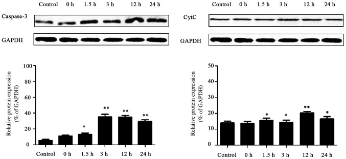
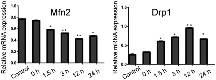
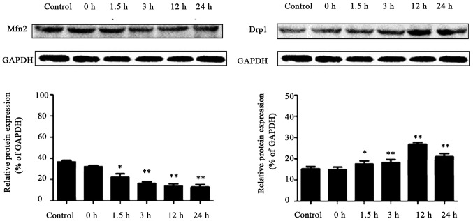
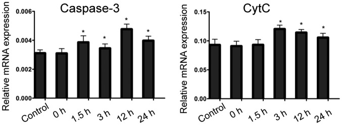
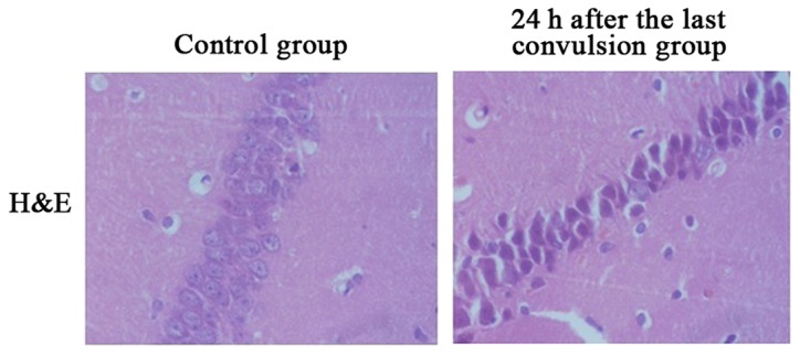
The aim of the present study was to establish a recurrent convulsion model during the developmental stage using inhalation of flurothyl, and to observe the relationship between the changes in mitochondrial function in hippocampal neurons and hippocampal neuronal apoptosis after recurrent convulsion. A total of 36 Sprague-Dawley male rats were selected and randomly divided into the control (NS) group and recurrent-seizure (RS) group for 0, 1.5, 3, 12 and 24 h. After the last seizure the rats were subdivided with 6 animals in each group. Rats in the seizure group inhaled flurothyl repeatedly to induce status convulsivus, 30 min once daily, for 7 consecutive days, while the same operation was conducted in the control group without inhalation of flurothyl. At each time-point after the last seizure, blood was taken from the heart, followed by decapitation and immediate removal of the brain. Half of the brain tissue was immediately fixed in 10% paraformaldehyde to prepare paraffin-embedded tissues for hematoxylin and eosin (H&E) histological staining. Hippocampus was taken from the other half of the brain and stored at -80°C. Changes in mitochondrial membrane potential (ΔΨm) in hippocampal neurons were detected by flow cytometer. Dynamic changes of mitochondrial fusion and division-related genes, mitochondrial fusion protein 2 (Mfn2) and dynamin-related protein 1 (Drp1), in the hippocampus after recurrent convulsion were observed using reverse transcription-polymerase chain reaction (RT-PCR)and western blot analysis. The expression of caspase-3 and cytochrome (Cyt c) was determined by RT-PCR and western blot analysis. After successful establishment of the recurrent convulsion model in rats during developmental stage using flurothyl, H&E staining results exhibited that in the CA1 region of hippocampus in the NS group, karyopyknosis occurred in nucleus that was stained to be brown and yellow, and the expression peak of apoptotic cells mainly existed at 24 h after the last convulsion. RT-PCR and western analysis revealed that apoptosis-related gene caspase-3 expression in the RS group was elevated at 1.5 h after the last convulsion, and lasted 24 h after convulsion. Detection results of mitochondrial ΔΨm revealed a significant reduction 1.5, 3 and 12 h after convulsion in hippocampal neurons of experimental rats, which reached the trough at 12 h, and rapidly increased after 24 h. The expression of Mfn2 mRNA in the RS group was significantly lower than that in the control group, while the expression of Drp1 mRNA in RS group was distinctly higher than that in the control group. RT-PCR and western blot analysis revealed that, mitochondrial apoptosis-related gene Cyt expression was increased at 3 h after the last convulsion, and lasted 24 h after convulsion. Correlation analysis showed that the changes in mitochondrial function were closely related to neuronal apoptosis. The results of the study show that apoptosis exists in the hippocampus of rats after recurrent convulsion, which is closely related to the changes in mitochondrial function.

https://cdn.ncbi.nlm.nih.gov/pmc/blobs/aa64/5995061/2f3396e581ad/etm-16-01-0127-g04.jpg
https://cdn.ncbi.nlm.nih.gov/pmc/blobs/aa64/5995061/78604173c809/etm-16-01-0127-g00.jpg
https://cdn.ncbi.nlm.nih.gov/pmc/blobs/aa64/5995061/4b9de746f943/etm-16-01-0127-g01.jpg
https://cdn.ncbi.nlm.nih.gov/pmc/blobs/aa64/5995061/e3ac09f8008f/etm-16-01-0127-g02.jpg
https://cdn.ncbi.nlm.nih.gov/pmc/blobs/aa64/5995061/02d9595b3e94/etm-16-01-0127-g03.jpg
https://cdn.ncbi.nlm.nih.gov/pmc/blobs/aa64/5995061/2f3396e581ad/etm-16-01-0127-g04.jpg
摘要

本研究旨在通过吸入三氟乙醚建立发育阶段反复惊厥模型,并观察反复惊厥后海马神经元线粒体功能变化与海马神经元凋亡之间的关系。选取36只Sprague-Dawley雄性大鼠,随机分为对照组(NS组)和反复惊厥组(RS组),分别在0、1.5、3、12和24小时进行观察。最后一次惊厥后,每组再细分6只动物。惊厥组大鼠每天吸入三氟乙醚30分钟,连续7天,反复诱导惊厥持续状态,对照组进行相同操作但不吸入三氟乙醚。在最后一次惊厥后的每个时间点,从心脏取血,随后断头并立即取出大脑。将一半脑组织立即固定于10%多聚甲醛中,制备石蜡包埋组织用于苏木精-伊红(H&E)组织学染色。从另一半脑组织中取出海马,储存于-80°C。采用流式细胞仪检测海马神经元线粒体膜电位(ΔΨm)变化。采用逆转录-聚合酶链反应(RT-PCR)和蛋白质免疫印迹分析观察反复惊厥后海马中线粒体融合与分裂相关基因、线粒体融合蛋白2(Mfn2)和动力相关蛋白1(Drp1)的动态变化。通过RT-PCR和蛋白质免疫印迹分析测定caspase-3和细胞色素C(Cyt c)的表达。利用三氟乙醚成功建立发育阶段大鼠反复惊厥模型后,H&E染色结果显示,NS组海马CA1区细胞核固缩,染成棕黄色,凋亡细胞表达高峰主要出现在最后一次惊厥后24小时。RT-PCR和蛋白质免疫分析显示,RS组凋亡相关基因caspase-3在最后一次惊厥后1.5小时表达升高,并在惊厥后持续24小时。线粒体ΔΨm检测结果显示,实验大鼠海马神经元在惊厥后1.5、3和12小时显著降低,在12小时达到谷底,24小时后迅速升高。RS组Mfn2 mRNA表达明显低于对照组,而RS组Drp1 mRNA表达明显高于对照组。RT-PCR和蛋白质免疫印迹分析显示,线粒体凋亡相关基因Cyt c在最后一次惊厥后3小时表达增加,并在惊厥后持续24小时。相关性分析表明,线粒体功能变化与神经元凋亡密切相关。研究结果表明,反复惊厥后大鼠海马存在凋亡,这与线粒体功能变化密切相关。

相似文献

1
Relationship between changes in mitochondrial function and hippocampal neuronal apoptosis after recurrent convulsion during developmental stage.发育阶段反复惊厥后线粒体功能变化与海马神经元凋亡的关系
Exp Ther Med. 2018 Jul;16(1):127-132. doi: 10.3892/etm.2018.6147. Epub 2018 May 10.
2
[Protective Effect of Acupuncture Serum Derived from Acute Convulsion Rats on Cultured Hip-pocampal Neurons with Seizure-like Discharges by Regulating Expression of Endoplasmic Reticulum Stress-inducible Molecular Chaperones].[急性惊厥大鼠针刺血清对具有癫痫样放电的培养海马神经元的保护作用:通过调节内质网应激诱导分子伴侣的表达]
Zhen Ci Yan Jiu. 2017 Apr 25;42(2):95-101.
3
Dynamic pattern of gene expression of ZnT-1, ZnT-3 and PRG-1 in rat brain following flurothyl-induced recurrent neonatal seizures.氟烷诱导新生鼠反复惊厥后大脑中 ZnT-1、ZnT-3 和 PRG-1 基因表达的动态变化。
Toxicol Lett. 2010 May 4;194(3):86-93. doi: 10.1016/j.toxlet.2010.02.008. Epub 2010 Feb 16.
4
Dynamic pattern of gene expression of ZnT-4, caspase-3, LC3, and PRG-3 in rat cerebral cortex following flurothyl-induced recurrent neonatal seizures.氟烷诱导新生鼠反复惊厥后大脑皮质 ZnT-4、caspase-3、LC3 和 PRG-3 基因表达的动态变化
Biol Trace Elem Res. 2011 Dec;143(3):1607-15. doi: 10.1007/s12011-011-8982-4. Epub 2011 Feb 1.
5
[Regulatory mechanism of MS275 on the p38 MAPK signaling pathway in rats with convulsion in the developmental stage].[MS275对发育阶段惊厥大鼠p38丝裂原活化蛋白激酶信号通路的调控机制]
Zhongguo Dang Dai Er Ke Za Zhi. 2020 Aug;22(8):909-915. doi: 10.7499/j.issn.1008-8830.2002167.
6
[Effects of propofol on expression of hippocampal survivin and Caspase-3 in newborn rats].[丙泊酚对新生大鼠海马生存素及半胱天冬酶-3表达的影响]
Zhonghua Er Ke Za Zhi. 2012 May;50(5):361-5.
7
ZnT-1, ZnT-3, CaMK II, PRG-1 expressions in hippocampus following neonatal seizure-induced cognitive deficit in rats.新生大鼠癫痫发作诱导认知缺陷后海马中锌转运体-1(ZnT-1)、锌转运体-3(ZnT-3)、钙/钙调蛋白依赖性蛋白激酶II(CaMK II)、富含脯氨酸的糖蛋白-1(PRG-1)的表达
Toxicol Lett. 2009 Feb 10;184(3):145-50. doi: 10.1016/j.toxlet.2008.11.003. Epub 2008 Nov 18.
8
[Effects of edaravone on the expression of interleukin-1beta, nuclear factor-kappaB and neuron apoptosis in juvenile rat hippocampus after status convulsion].依达拉奉对惊厥持续状态幼鼠海马白细胞介素-1β、核因子-κB表达及神经元凋亡的影响
Zhonghua Er Ke Za Zhi. 2009 Aug;47(8):575-80.
9
[Effect of duration of convulsion state on neuronal apoptosis and early apoptotic events in hippocampus of rats].[惊厥状态持续时间对大鼠海马神经元凋亡及早期凋亡事件的影响]
Zhongguo Wei Zhong Bing Ji Jiu Yi Xue. 2007 Jun;19(6):343-5.
10
[Effects of mitochondrial division inhibitor on neurological function and neuronal apoptosis in rats after cardiopulmonary resuscitation].线粒体分裂抑制剂对心肺复苏后大鼠神经功能及神经元凋亡的影响
Zhonghua Wei Zhong Bing Ji Jiu Yi Xue. 2015 Dec;27(12):950-4. doi: 10.3760/cma.j.issn.2095-4352.2015.12.002.

引用本文的文献

1
Circadian Clock Genes REV-ERBs Inhibits Granulosa Cells Apoptosis by Regulating Mitochondrial Biogenesis and Autophagy in Polycystic Ovary Syndrome.昼夜节律时钟基因REV-ERBs通过调节多囊卵巢综合征中颗粒细胞的线粒体生物合成和自噬来抑制颗粒细胞凋亡。
Front Cell Dev Biol. 2021 Aug 5;9:658112. doi: 10.3389/fcell.2021.658112. eCollection 2021.
2
Proteomics for Studying the Effects of Ketogenic Diet Against Lithium Chloride/Pilocarpine Induced Epilepsy in Rats.蛋白质组学用于研究生酮饮食对氯化锂/毛果芸香碱诱导的大鼠癫痫的影响。
Front Neurosci. 2020 Sep 29;14:562853. doi: 10.3389/fnins.2020.562853. eCollection 2020.
3
Targeted Antitumor Mechanism of C-PC/CMC-CD55sp Nanospheres in HeLa Cervical Cancer Cells.

本文引用的文献

1
Adiponectin partially rescues high glucose/high fat-induced impairment of mitochondrial biogenesis and function in a PGC-1α dependent manner.脂联素以依赖于PGC-1α的方式部分挽救高糖/高脂诱导的线粒体生物合成和功能损伤。
Eur Rev Med Pharmacol Sci. 2017 Jan;21(3):590-599.
2
2014 AHA/ACC/HRS guideline for the management of patients with atrial fibrillation: a report of the American College of Cardiology/American Heart Association Task Force on Practice Guidelines and the Heart Rhythm Society.2014年美国心脏协会/美国心脏病学会/心律学会心房颤动患者管理指南:美国心脏病学会/美国心脏协会实践指南工作组及心律学会的报告
J Am Coll Cardiol. 2014 Dec 2;64(21):e1-76. doi: 10.1016/j.jacc.2014.03.022. Epub 2014 Mar 28.
3
C-PC/CMC-CD55sp纳米球对HeLa宫颈癌细胞的靶向抗肿瘤机制
Front Pharmacol. 2020 Jun 18;11:906. doi: 10.3389/fphar.2020.00906. eCollection 2020.
Cognitive outcome of status epilepticus in children.
儿童癫痫持续状态的认知结局
Epilepsy Res Treat. 2012;2012:984124. doi: 10.1155/2012/984124. Epub 2012 Aug 13.
4
miR-181 targets multiple Bcl-2 family members and influences apoptosis and mitochondrial function in astrocytes.miR-181 靶向多个 Bcl-2 家族成员并影响星形胶质细胞中的细胞凋亡和线粒体功能。
Mitochondrion. 2012 Mar;12(2):213-9. doi: 10.1016/j.mito.2011.09.001. Epub 2011 Sep 17.
5
Developmental outcome after a single episode of status epilepticus.单次癫痫持续状态后的发育结局。
Epilepsy Behav. 2011 Aug;21(4):430-6. doi: 10.1016/j.yebeh.2011.05.009. Epub 2011 Jun 25.
6
An excitatory loop with astrocytes contributes to drive neurons to seizure threshold.星形胶质细胞的兴奋性循环有助于驱动神经元达到癫痫发作阈值。
PLoS Biol. 2010 Apr 13;8(4):e1000352. doi: 10.1371/journal.pbio.1000352.
7
Treatment of community-onset, childhood convulsive status epilepticus: a prospective, population-based study.社区获得性儿童惊厥性癫痫持续状态的治疗:一项基于人群的前瞻性研究。
Lancet Neurol. 2008 Aug;7(8):696-703. doi: 10.1016/S1474-4422(08)70141-8. Epub 2008 Jul 2.
8
Three-dimensional structure of the apoptosome: implications for assembly, procaspase-9 binding, and activation.凋亡小体的三维结构:对组装、前半胱天冬酶-9结合及激活的意义
Mol Cell. 2002 Feb;9(2):423-32. doi: 10.1016/s1097-2765(02)00442-2.
9
Mitochondria and calcium: from cell signalling to cell death.线粒体与钙:从细胞信号传导到细胞死亡
J Physiol. 2000 Nov 15;529 Pt 1(Pt 1):57-68. doi: 10.1111/j.1469-7793.2000.00057.x.
10
Corpse clearance defines the meaning of cell death.尸体清除定义了细胞死亡的意义。
Nature. 2000 Oct 12;407(6805):784-8. doi: 10.1038/35037722.