Second Department of Surgery, Hamamatsu University School of Medicine, Hamamatsu, Japan.
Department of Cellular and Molecular Anatomy, International Mass Imaging Center, Hamamatsu University School of Medicine, Hamamatsu, Japan.
Cancer Sci. 2018 Aug;109(8):2401-2411. doi: 10.1111/cas.13659. Epub 2018 Jul 7.
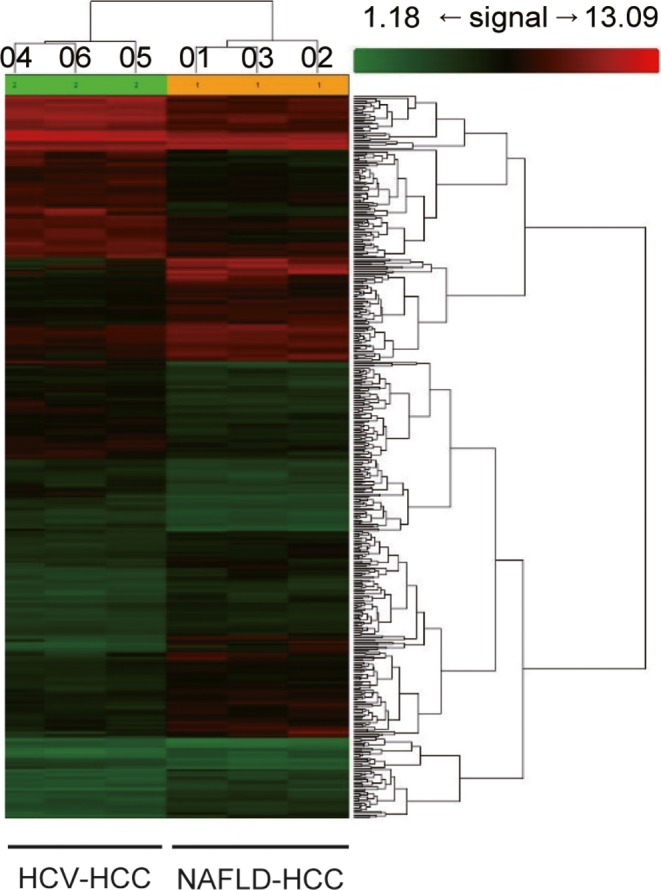
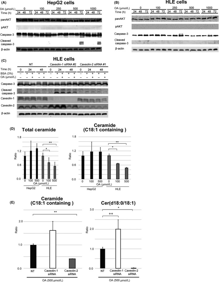
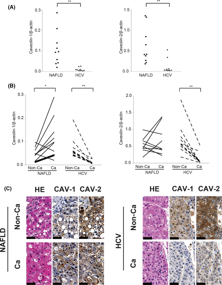
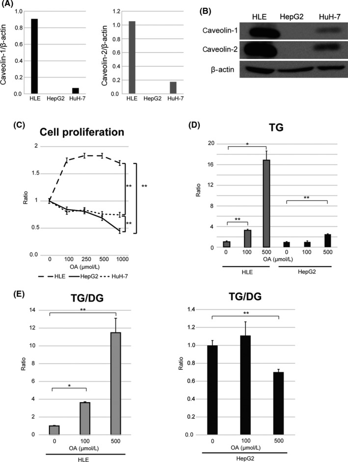
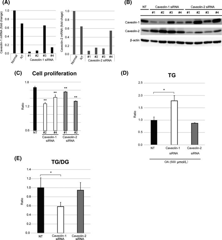
The molecular features of hepatocellular carcinoma arising from non-alcoholic fatty liver disease (NAFLD-HCC) are not well known. In this study, we investigated the mechanism by which NAFLD-HCC survives in a fat-rich environment. We found that caveolin (CAV)-1 was overexpressed in clinical specimens from NAFLD-HCC patients. HepG2, HLE, and HuH-7 HCC cell lines showed decreased proliferation in the presence of the saturated fatty acids palmitic acid and stearic acid, although only HLE cells expressed high levels of CAV-1. HLE cells treated with oleic acid (OA) showed robust proliferation, whereas CAV-null HepG2 cells showed reduced proliferation and increased apoptosis. CAV-1 knockdown in HLE cells attenuated the OA-induced increase in proliferation and enhanced apoptosis. Liquid chromatography-tandem mass spectrometry analysis revealed that the levels of OA-containing ceramide, a pro-apoptotic factor, were higher in HepG2 and CAV-1-deficient HLE cells than in HLE cells, suggesting that CAV-1 inhibits apoptosis by decreasing the level of OA-containing ceramide. These results indicate that CAV-1 is important for NAFLD-HCC survival in fatty acid-rich environments and is a potential therapeutic target.
非酒精性脂肪性肝病(NAFLD)相关肝细胞癌(HCC)的分子特征尚不清楚。本研究旨在探讨富含脂肪的环境中 NAFLD-HCC 存活的机制。我们发现,在 NAFLD-HCC 患者的临床标本中,窖蛋白(CAV)-1 过表达。HepG2、HLE 和 HuH-7 HCC 细胞系在饱和脂肪酸棕榈酸和硬脂酸存在下增殖减少,尽管只有 HLE 细胞表达高水平的 CAV-1。用油酸(OA)处理的 HLE 细胞表现出旺盛的增殖,而 CAV 缺失的 HepG2 细胞增殖减少,凋亡增加。HLE 细胞中 CAV-1 的敲低可减弱 OA 诱导的增殖增加并增强凋亡。液质联用分析显示,含 OA 的神经酰胺(一种促凋亡因子)的水平在 HepG2 和 CAV-1 缺陷型 HLE 细胞中高于 HLE 细胞,表明 CAV-1 通过降低含 OA 的神经酰胺水平抑制凋亡。这些结果表明,CAV-1 对于富含脂肪酸的环境中 NAFLD-HCC 的存活很重要,是一个潜在的治疗靶点。