Department of Biomedical Engineering, University of Virginia, Charlottesville, VA.
Theranostics. 2018 Apr 20;8(11):2988-2991. doi: 10.7150/thno.26025. eCollection 2018.
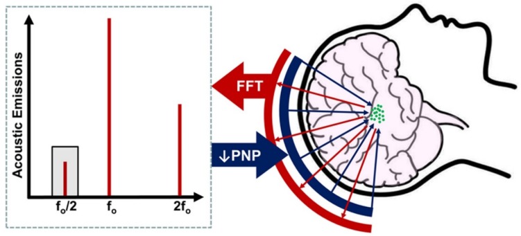
Non-invasive drug and gene delivery to the brain to treat central nervous system pathologies has long been inhibited by the blood-brain barrier. The activation of microbubbles with focused ultrasound has emerged as a promising non-invasive approach to circumvent this obstacle, by transiently disrupting the blood-brain barrier and permitting passage of systemically administered therapeutics into the tissue. Clinical trials are underway to evaluate the safety of this technique; however, concerns remain regarding the potential for the treatment to induce sterile inflammation or petechiae. In this issue of , Jones et al.[1] address these concerns through the development of an advanced three-dimensional imaging system for monitoring acoustic emissions from oscillating microbubbles. When subharmonic emissions are detected with this system, focused ultrasound pressure is reduced by 50% for the remainder of the treatment. This serves to transiently open the blood-brain barrier without generating adverse effects. While the ideal configuration of the transducer array for treatment and monitoring still presents an area for further optimization, the approach indicates that the acoustic signature of microbubble behavior within the skull can be used to ensure safe and effective blood-brain barrier opening using focused ultrasound.
长期以来,由于血脑屏障的存在,向大脑输送非侵入性药物和基因来治疗中枢神经系统疾病一直受到限制。利用聚焦超声激活微泡已成为一种很有前途的非侵入性方法,可以暂时破坏血脑屏障,使系统给予的治疗药物进入组织。目前正在进行临床试验以评估该技术的安全性;然而,人们仍然担心该治疗方法会引起无菌性炎症或瘀点。在本期 杂志中,Jones 等人[1]通过开发一种用于监测微泡振荡声发射的先进的三维成像系统来解决这些问题。当使用该系统检测到次谐波发射时,在治疗的剩余时间内,聚焦超声的压力降低 50%。这可以暂时打开血脑屏障,而不会产生不良反应。虽然治疗和监测用换能器阵列的理想配置仍然是一个需要进一步优化的领域,但该方法表明,可以利用颅骨内微泡行为的声特征来确保使用聚焦超声安全有效地打开血脑屏障。