• 文献检索
  • 文档翻译
  • 深度研究
  • 学术资讯
  • Suppr Zotero 插件Zotero 插件
  • 邀请有礼
  • 套餐&价格
  • 历史记录
应用&插件
Suppr Zotero 插件Zotero 插件浏览器插件Mac 客户端Windows 客户端微信小程序
定价
高级版会员购买积分包购买API积分包
服务
文献检索文档翻译深度研究API 文档MCP 服务
关于我们
关于 Suppr公司介绍联系我们用户协议隐私条款
关注我们

Suppr 超能文献

核心技术专利:CN118964589B侵权必究
粤ICP备2023148730 号-1Suppr @ 2026

文献检索

告别复杂PubMed语法,用中文像聊天一样搜索,搜遍4000万医学文献。AI智能推荐,让科研检索更轻松。

立即免费搜索

文件翻译

保留排版,准确专业,支持PDF/Word/PPT等文件格式,支持 12+语言互译。

免费翻译文档

深度研究

AI帮你快速写综述,25分钟生成高质量综述,智能提取关键信息,辅助科研写作。

立即免费体验

室性期前收缩的激活后恢复间期成像。

Activation recovery interval imaging of premature ventricular contraction.

机构信息

Department of Biomedical Engineering, University of Minnesota, Minneapolis, MN, United States of America.

Department of Cardiology, Shanghai Ruijin Hospital, Shanghai, China.

出版信息

PLoS One. 2018 Jun 15;13(6):e0196916. doi: 10.1371/journal.pone.0196916. eCollection 2018.

DOI:10.1371/journal.pone.0196916
PMID:29906289
原文链接:https://pmc.ncbi.nlm.nih.gov/articles/PMC6003683/
Abstract

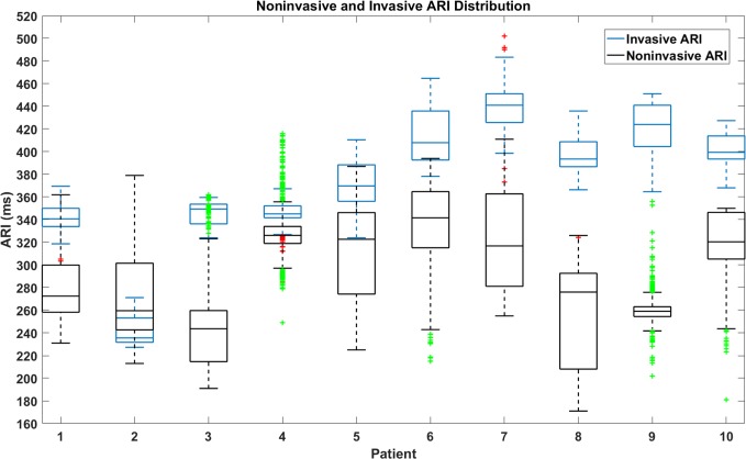
Dispersion of ventricular repolarization due to abnormal activation contributes to the susceptibility to cardiac arrhythmias. However, the global pattern of repolarization is difficult to assess clinically. Activation recovery interval (ARI) has been used to understand the properties of ventricular repolarization. In this study, we developed an ARI imaging technique to noninvasively reconstruct three-dimensional (3D) ARI maps in 10 premature ventricular contraction (PVC) patients and evaluated the results with the endocardial ARI maps recorded by a clinical navigation system (CARTO). From the analysis results of a total of 100 PVC beats in 10 patients, the average correlation coefficient is 0.86±0.05 and the average relative error is 0.06±0.03. The average localization error is 4.5±2.3 mm between the longest ARI sites in 3D ARI maps and those in CARTO endocardial ARI maps. The present results suggest that ARI imaging could serve as an alternative of evaluating global pattern of ventricular repolarization noninvasively and could assist in the future investigation of the relationship between global repolarization dispersion and the susceptibility to cardiac arrhythmias.

摘要

心室复极的离散性是由于异常激活导致的,这会增加心脏心律失常的易感性。然而,整体复极模式在临床上很难评估。激活后恢复间期(ARI)已被用于了解心室复极的特性。在这项研究中,我们开发了一种 ARI 成像技术,以非侵入性地重建 10 名室性早搏(PVC)患者的三维(3D)ARI 图谱,并将结果与临床导航系统(CARTO)记录的心内膜 ARI 图谱进行比较。通过对 10 名患者的 100 个 PVC 搏动的分析结果,平均相关系数为 0.86±0.05,平均相对误差为 0.06±0.03。在 3D ARI 图谱和 CARTO 心内膜 ARI 图谱中最长 ARI 部位之间的平均定位误差为 4.5±2.3mm。这些结果表明,ARI 成像可以作为评估心室复极整体模式的一种替代方法,有助于未来研究整体复极离散性与心脏心律失常易感性之间的关系。

https://cdn.ncbi.nlm.nih.gov/pmc/blobs/95f4/6003683/f23a0251b576/pone.0196916.g007.jpg
https://cdn.ncbi.nlm.nih.gov/pmc/blobs/95f4/6003683/8a2c71ec77f8/pone.0196916.g001.jpg
https://cdn.ncbi.nlm.nih.gov/pmc/blobs/95f4/6003683/a1689b4ec638/pone.0196916.g002.jpg
https://cdn.ncbi.nlm.nih.gov/pmc/blobs/95f4/6003683/ed5d536f2aae/pone.0196916.g003.jpg
https://cdn.ncbi.nlm.nih.gov/pmc/blobs/95f4/6003683/9aabebbdc117/pone.0196916.g004.jpg
https://cdn.ncbi.nlm.nih.gov/pmc/blobs/95f4/6003683/b7acea865494/pone.0196916.g005.jpg
https://cdn.ncbi.nlm.nih.gov/pmc/blobs/95f4/6003683/a3ff1e022923/pone.0196916.g006.jpg
https://cdn.ncbi.nlm.nih.gov/pmc/blobs/95f4/6003683/f23a0251b576/pone.0196916.g007.jpg

相似文献

1
Activation recovery interval imaging of premature ventricular contraction.室性期前收缩的激活后恢复间期成像。
PLoS One. 2018 Jun 15;13(6):e0196916. doi: 10.1371/journal.pone.0196916. eCollection 2018.
2
Noninvasive reconstruction of the three-dimensional ventricular activation sequence during pacing and ventricular tachycardia in the canine heart.非侵入性重建犬心起搏和室速时的三维心室激动顺序。
Am J Physiol Heart Circ Physiol. 2012 Jan 1;302(1):H244-52. doi: 10.1152/ajpheart.00618.2011. Epub 2011 Oct 7.
3
Three-Dimensional Noninvasive Imaging of Ventricular Arrhythmias in Patients With Premature Ventricular Contractions.三维非侵入性成像在室性早搏患者中的心律失常。
IEEE Trans Biomed Eng. 2018 Jul;65(7):1495-1503. doi: 10.1109/TBME.2017.2758369. Epub 2017 Oct 2.
4
Noninvasive cardiac activation imaging of ventricular arrhythmias during drug-induced QT prolongation in the rabbit heart.兔心药物诱导 QT 间期延长时室性心律失常的无创性心脏激活成像。
Heart Rhythm. 2013 Oct;10(10):1509-15. doi: 10.1016/j.hrthm.2013.06.010. Epub 2013 Jun 14.
5
Modulated dispersion of activation and repolarization by premature beats in patients with cardiomyopathy at risk of sudden death.**译文**:致心律失常性右室心肌病患者中,通过期前收缩调制的激活和复极的离散度。
Am J Physiol Heart Circ Physiol. 2011 Jun;300(6):H2221-9. doi: 10.1152/ajpheart.01050.2010. Epub 2011 Mar 25.
6
Activation recovery time measurements in evaluation of global sequence and dispersion of ventricular repolarization.在评估心室复极的整体序列和离散度时进行激活恢复时间测量。
J Electrocardiol. 2005 Jan;38(1):28-35. doi: 10.1016/j.jelectrocard.2004.09.010.
7
Noninvasive Mapping of Premature Ventricular Contractions by Merging Magnetocardiography and Computed Tomography.应用心磁图和计算机断层扫描技术无创性定位室性期前收缩。
JACC Clin Electrophysiol. 2019 Oct;5(10):1144-1157. doi: 10.1016/j.jacep.2019.06.010. Epub 2019 Jul 31.
8
Increased ventricular repolarization heterogeneity in patients with ventricular arrhythmia vulnerability and cardiomyopathy: a human in vivo study.室性心律失常易感性和心肌病患者心室复极异质性增加:一项人体体内研究。
Am J Physiol Heart Circ Physiol. 2006 Jan;290(1):H79-86. doi: 10.1152/ajpheart.00648.2005. Epub 2005 Aug 19.
9
Determination of human ventricular repolarization by noncontact mapping: validation with monophasic action potential recordings.通过非接触式标测确定人体心室复极:单相动作电位记录验证
Circulation. 2004 Sep 14;110(11):1343-50. doi: 10.1161/01.CIR.0000141734.43393.BE. Epub 2004 Sep 7.
10
Arrhythmogenesis of T wave alternans associated with surface QRS complex alternans and the role of ventricular prematurity: observations from a canine model of LQT3 syndrome.与体表QRS波群交替相关的T波交替的心律失常发生机制及室性早搏的作用:来自长QT3综合征犬模型的观察
J Cardiovasc Electrophysiol. 2002 Jun;13(6):599-604. doi: 10.1046/j.1540-8167.2002.00599.x.

引用本文的文献

1
Developing cardiac digital twin populations powered by machine learning provides electrophysiological insights in conduction and repolarization.利用机器学习开发心脏数字孪生群体可提供有关传导和复极化的电生理见解。
Nat Cardiovasc Res. 2025 May;4(5):624-636. doi: 10.1038/s44161-025-00650-0. Epub 2025 May 16.
2
Conduction Velocity Dispersion Predicts Postinfarct Ventricular Tachycardia Circuit Sites and Associates With Lipomatous Metaplasia.传导速度离散度预测梗死后性室性心动过速的折返环部位,并与脂肪化生相关。
JACC Clin Electrophysiol. 2023 Aug;9(8 Pt 2):1464-1474. doi: 10.1016/j.jacep.2023.05.013. Epub 2023 May 18.

本文引用的文献

1
In Vivo Validation of Electrocardiographic Imaging.体内心电图成像的验证。
JACC Clin Electrophysiol. 2017 Mar;3(3):232-242. doi: 10.1016/j.jacep.2016.11.012. Epub 2017 Feb 1.
2
Three-Dimensional Noninvasive Imaging of Ventricular Arrhythmias in Patients With Premature Ventricular Contractions.三维非侵入性成像在室性早搏患者中的心律失常。
IEEE Trans Biomed Eng. 2018 Jul;65(7):1495-1503. doi: 10.1109/TBME.2017.2758369. Epub 2017 Oct 2.
3
New Insights in Cardiac Calcium Handling and Excitation-Contraction Coupling.
心脏钙处理和兴奋-收缩偶联的新见解。
Adv Exp Med Biol. 2018;1067:373-385. doi: 10.1007/5584_2017_106.
4
Noninvasive Personalization of a Cardiac Electrophysiology Model From Body Surface Potential Mapping.基于体表电位标测的心脏电生理模型无创个性化构建
IEEE Trans Biomed Eng. 2017 Sep;64(9):2206-2218. doi: 10.1109/TBME.2016.2629849. Epub 2016 Nov 16.
5
Noninvasive Imaging of High-Frequency Drivers and Reconstruction of Global Dominant Frequency Maps in Patients With Paroxysmal and Persistent Atrial Fibrillation.阵发性和持续性心房颤动患者高频驱动因素的无创成像及整体主导频率图的重建
IEEE Trans Biomed Eng. 2016 Jun;63(6):1333-1340. doi: 10.1109/TBME.2016.2553641. Epub 2016 Apr 13.
6
Temporal Sparse Promoting Three Dimensional Imaging of Cardiac Activation.时间稀疏促进心脏激活的三维成像
IEEE Trans Med Imaging. 2015 Nov;34(11):2309-19. doi: 10.1109/TMI.2015.2429134. Epub 2015 May 4.
7
Sensitivity of Noninvasive Cardiac Electrophysiological Imaging to Variations in Personalized Anatomical Modeling.非侵入性心脏电生理成像对个性化解剖模型变化的敏感性。
IEEE Trans Biomed Eng. 2015 Jun;62(6):1563-75. doi: 10.1109/TBME.2015.2395387. Epub 2015 Jan 21.
8
Imaging cardiac activation sequence during ventricular tachycardia in a canine model of nonischemic heart failure.在非缺血性心力衰竭犬模型中对室性心动过速期间的心脏激活序列进行成像。
Am J Physiol Heart Circ Physiol. 2015 Jan 15;308(2):H108-14. doi: 10.1152/ajpheart.00196.2014. Epub 2014 Nov 21.
9
Measurement bias in activation-recovery intervals from unipolar electrograms.单极电图激活-恢复间期的测量偏差
Am J Physiol Heart Circ Physiol. 2015 Feb 15;308(4):H331-8. doi: 10.1152/ajpheart.00478.2014. Epub 2014 Nov 14.
10
Assessment of regularization techniques for electrocardiographic imaging.心电图成像正则化技术评估
J Electrocardiol. 2014 Jan-Feb;47(1):20-8. doi: 10.1016/j.jelectrocard.2013.10.004. Epub 2013 Oct 17.