• 文献检索
  • 文档翻译
  • 深度研究
  • 学术资讯
  • Suppr Zotero 插件Zotero 插件
  • 邀请有礼
  • 套餐&价格
  • 历史记录
应用&插件
Suppr Zotero 插件Zotero 插件浏览器插件Mac 客户端Windows 客户端微信小程序
定价
高级版会员购买积分包购买API积分包
服务
文献检索文档翻译深度研究API 文档MCP 服务
关于我们
关于 Suppr公司介绍联系我们用户协议隐私条款
关注我们

Suppr 超能文献

核心技术专利:CN118964589B侵权必究
粤ICP备2023148730 号-1Suppr @ 2026

文献检索

告别复杂PubMed语法,用中文像聊天一样搜索,搜遍4000万医学文献。AI智能推荐,让科研检索更轻松。

立即免费搜索

文件翻译

保留排版,准确专业,支持PDF/Word/PPT等文件格式,支持 12+语言互译。

免费翻译文档

深度研究

AI帮你快速写综述,25分钟生成高质量综述,智能提取关键信息,辅助科研写作。

立即免费体验

低剂量 Evans 蓝染料在光血栓性脑卒中模型中的近红外荧光成像。

Low-Dose Evans Blue Dye for Near-Infrared Fluorescence Imaging in Photothrombotic Stroke Model.

机构信息

Department of Forensic Medicine.

Department of Premedical Program, School of Medicine, Chosun University, Gwangju 61452, South Korea.

出版信息

Int J Med Sci. 2018 Apr 27;15(7):696-702. doi: 10.7150/ijms.24257. eCollection 2018.

DOI:10.7150/ijms.24257
PMID:29910674
原文链接:https://pmc.ncbi.nlm.nih.gov/articles/PMC6001419/
Abstract

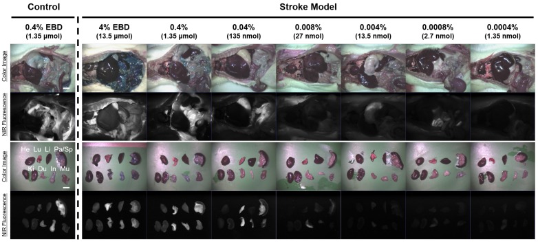
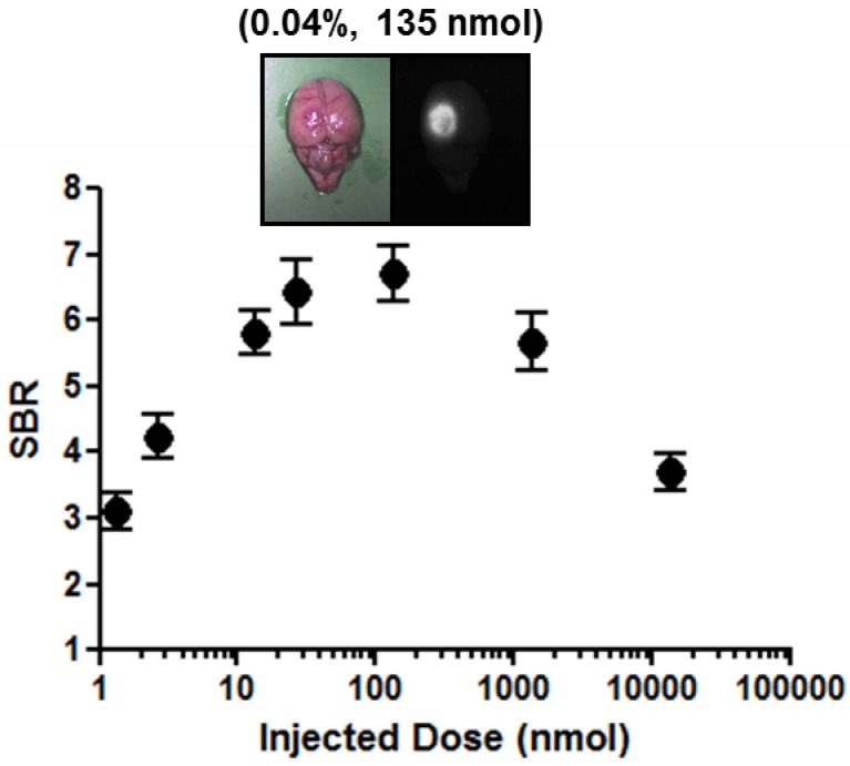
Evans blue dye (EBD) is the most common indicator to analyze the extent of blood-brain barrier (BBB) breakdown in several neurological disease models. However, the high-dose of EBD (51.9 mg/kg) is usually required for visualization of blue color by the human eye that brings potential safety issues. To solve this problem, low-dose of EBD was applied for the near-infrared (NIR) fluorescence-assisted quantitation of BBB breakdown in photothrombotic stoke model. Animals were allocated to seven dose groups ranging from 1.35 nmol (5.19 μg/kg) to 13.5 μmol (51.9 mg/kg) EBD. EBD was undetectable in the non-ischemic brain tissue, and the fluorescence signals in the infarcted hemisphere seemed proportional to the injected dose in the dose range. Although the maximum fluorescence signals in brain tissue were obtained with the injections of 1.35 nmol ~ 13.5 μmol EBD, the background signals in the neighboring brain tissues were significantly increased as well. Since the high concentration of EBD is necessary for color-based identification of the infarcted lesion in brain tissues, even 10-fold diluted could not be distinguished visually by naked eye. NIR fluorescence-assisted method could potentially provide new opportunities to study BBB leakage just using small amount of EBD in different pathological conditions and to test the efficacy of various therapeutic strategies to protect the BBB.

摘要

伊文思蓝染料(EBD)是分析几种神经疾病模型中血脑屏障(BBB)破裂程度的最常用指示剂。然而,为了用肉眼观察到蓝色,通常需要使用 EBD 的高剂量(51.9mg/kg),这带来了潜在的安全问题。为了解决这个问题,在光血栓性中风模型中,使用低剂量的 EBD 进行近红外(NIR)荧光辅助定量 BBB 破裂。动物被分为七个剂量组,EBD 剂量范围从 1.35nmol(5.19μg/kg)到 13.5μmol(51.9mg/kg)。非缺血性脑组织中无法检测到 EBD,并且梗塞侧半球的荧光信号似乎与注射剂量在剂量范围内呈比例关系。尽管在注射 1.35nmol 到 13.5μmol EBD 时,脑组织中获得了最大的荧光信号,但邻近脑组织中的背景信号也显著增加。由于高浓度的 EBD 对于基于颜色的脑组织梗塞病变的识别是必要的,即使是 10 倍稀释也无法用肉眼区分。NIR 荧光辅助方法有可能为研究不同病理条件下 BBB 渗漏提供新的机会,仅使用少量的 EBD,并测试各种保护 BBB 的治疗策略的疗效。

https://cdn.ncbi.nlm.nih.gov/pmc/blobs/7d4a/6001419/aff916ee0536/ijmsv15p0696g004.jpg
https://cdn.ncbi.nlm.nih.gov/pmc/blobs/7d4a/6001419/fac5a720cea5/ijmsv15p0696g001.jpg
https://cdn.ncbi.nlm.nih.gov/pmc/blobs/7d4a/6001419/84a5b19823ea/ijmsv15p0696g002.jpg
https://cdn.ncbi.nlm.nih.gov/pmc/blobs/7d4a/6001419/78442253f859/ijmsv15p0696g003.jpg
https://cdn.ncbi.nlm.nih.gov/pmc/blobs/7d4a/6001419/aff916ee0536/ijmsv15p0696g004.jpg

相似文献

1
Low-Dose Evans Blue Dye for Near-Infrared Fluorescence Imaging in Photothrombotic Stroke Model.低剂量 Evans 蓝染料在光血栓性脑卒中模型中的近红外荧光成像。
Int J Med Sci. 2018 Apr 27;15(7):696-702. doi: 10.7150/ijms.24257. eCollection 2018.
2
ZW800-1 for Assessment of Blood-Brain Barrier Disruption in a Photothrombotic Stroke Model.ZW800-1 用于评估光血栓性中风模型中的血脑屏障破坏。
Int J Med Sci. 2017 Nov 2;14(13):1430-1435. doi: 10.7150/ijms.22294. eCollection 2017.
3
Vascular delivery of intraperitoneal Evans blue dye into the blood-brain barrier-intact and disrupted rat brains.将腹腔内伊文思蓝染料经血管输送到血脑屏障完整和破坏的大鼠脑内。
Neuroreport. 2018 Aug 1;29(11):924-931. doi: 10.1097/WNR.0000000000001052.
4
Increase in Evans blue dye extravasation into the brain in the late developmental stage.发育后期伊文思蓝染料向脑内的外渗增加。
Neuroreport. 2012 Aug 22;23(12):699-701. doi: 10.1097/WNR.0b013e3283556dcc.
5
A new micro-computed tomography-based high-resolution blood-brain barrier imaging technique to study ischemic stroke.一种基于新的微计算机断层扫描的高分辨率血脑屏障成像技术,用于研究缺血性中风。
Stroke. 2014 Aug;45(8):2480-4. doi: 10.1161/STROKEAHA.114.006297. Epub 2014 Jul 10.
6
Optimization of Evans blue quantitation in limited rat tissue samples.有限大鼠组织样本中伊文思蓝定量的优化
Sci Rep. 2014 Oct 10;4:6588. doi: 10.1038/srep06588.
7
Permeability of the blood-brain barrier within rat intrastriatal transplants assessed by simultaneous systemic injection of horseradish peroxidase and Evans blue dye.通过同时全身注射辣根过氧化物酶和伊文思蓝染料评估大鼠纹状体内移植体中血脑屏障的通透性。
Exp Neurol. 1994 Jun;127(2):245-52. doi: 10.1006/exnr.1994.1100.
8
In vivo monitoring blood-brain barrier permeability using spectral imaging through optical clearing skull window.利用光学透明颅骨窗的光谱成象技术进行活体监测血脑屏障通透性。
J Biophotonics. 2019 Apr;12(4):e201800330. doi: 10.1002/jbio.201800330. Epub 2018 Dec 20.
9
Intravenous mannitol does not increase blood-brain barrier permeability to inert dyes in the adult rat forebrain.静脉注射甘露醇不会增加成年大鼠前脑血脑屏障对惰性染料的通透性。
Neuroreport. 2013 Apr 17;24(6):303-7. doi: 10.1097/WNR.0b013e32835f8acb.
10
Rodent stroke induced by photochemical occlusion of proximal middle cerebral artery: evolution monitored with MR imaging and histopathology.大脑中动脉近端光化学闭塞诱导的啮齿动物中风:通过磁共振成像和组织病理学监测其演变
Eur J Radiol. 2007 Jul;63(1):68-75. doi: 10.1016/j.ejrad.2007.01.005. Epub 2007 Mar 6.

引用本文的文献

1
Evaluation of Blood-Brain Barrier Disruption Using Low- and High-Molecular-Weight Complexes in a Single Brain Sample in a Rat Traumatic Brain Injury Model: Comparison to an Established Magnetic Resonance Imaging Technique.在大鼠创伤性脑损伤模型中,使用单一脑样本中的低和高分子量复合物评估血脑屏障破坏:与一种已建立的磁共振成像技术的比较。
Int J Mol Sci. 2024 Oct 19;25(20):11241. doi: 10.3390/ijms252011241.
2
Localized Increased Permeability of Blood-Brain Barrier for Antibody Conjugates in the Cuprizone Model of Demyelination.在脱髓鞘的 Cuprizone 模型中,抗体缀合物的血脑屏障通透性增加。
Int J Mol Sci. 2023 Aug 11;24(16):12688. doi: 10.3390/ijms241612688.
3

本文引用的文献

1
ZW800-1 for Assessment of Blood-Brain Barrier Disruption in a Photothrombotic Stroke Model.ZW800-1 用于评估光血栓性中风模型中的血脑屏障破坏。
Int J Med Sci. 2017 Nov 2;14(13):1430-1435. doi: 10.7150/ijms.22294. eCollection 2017.
2
Real-time imaging of metastatic bone tumors with a targeted near-infrared fluorophore.使用靶向近红外荧光团对转移性骨肿瘤进行实时成像。
Oncotarget. 2017 Aug 11;8(39):65770-65777. doi: 10.18632/oncotarget.20187. eCollection 2017 Sep 12.
3
Structure-Inherent Targeting of Near-Infrared Fluorophores for Image-Guided Surgery.
Endothelial Specific Deletion of Autotaxin Improves Stroke Outcomes.
内皮细胞特异性敲除 ATX 可改善脑卒中结局。
Cells. 2023 Feb 3;12(3):511. doi: 10.3390/cells12030511.
4
The ATX-LPA Axis Regulates Vascular Permeability during Cerebral Ischemic-Reperfusion.ATX-LPA 轴在脑缺血再灌注期间调节血管通透性。
Int J Mol Sci. 2022 Apr 8;23(8):4138. doi: 10.3390/ijms23084138.
5
Disrupted Blood-Brain Barrier and Mitochondrial Impairment by Autotaxin-Lysophosphatidic Acid Axis in Postischemic Stroke.缺血性脑卒中后自分泌酶-溶血磷脂酸轴导致血脑屏障破坏和线粒体损伤。
J Am Heart Assoc. 2021 Sep 21;10(18):e021511. doi: 10.1161/JAHA.121.021511. Epub 2021 Sep 13.
6
Circulating tight-junction proteins are potential biomarkers for blood-brain barrier function in a model of neonatal hypoxic/ischemic brain injury.循环紧密连接蛋白是新生儿缺氧缺血性脑损伤模型中血脑屏障功能的潜在生物标志物。
Fluids Barriers CNS. 2021 Feb 10;18(1):7. doi: 10.1186/s12987-021-00240-9.
7
Enhanced spinal neuronal responses as a mechanism for increased number and size of active acupoints in visceral hyperalgesia.增强的脊髓神经元反应是内脏痛觉过敏中活跃穴位数量和大小增加的机制。
Sci Rep. 2020 Jun 25;10(1):10312. doi: 10.1038/s41598-020-67242-9.
8
Bioluminescence and near-infrared fluorescence imaging for detection of metastatic bone tumors.生物发光和近红外荧光成像用于检测转移性骨肿瘤。
Lasers Med Sci. 2020 Feb;35(1):115-120. doi: 10.1007/s10103-019-02801-9. Epub 2019 Jun 1.
用于图像引导手术的近红外荧光团的结构固有靶向
Chonnam Med J. 2017 May;53(2):95-102. doi: 10.4068/cmj.2017.53.2.95. Epub 2017 May 25.
4
Overexpression of caveolin-1 attenuates brain edema by inhibiting tight junction degradation.小窝蛋白-1的过表达通过抑制紧密连接的降解来减轻脑水肿。
Oncotarget. 2016 Oct 18;7(42):67857-67867. doi: 10.18632/oncotarget.12346.
5
Biocompatible Red Fluorescent Organic Nanoparticles with Tunable Size and Aggregation-Induced Emission for Evaluation of Blood-Brain Barrier Damage.具有可调尺寸和聚集诱导发射的生物相容红色荧光有机纳米粒子用于评估血脑屏障损伤。
Adv Mater. 2016 Oct;28(39):8760-8765. doi: 10.1002/adma.201601191. Epub 2016 Aug 11.
6
Neuroprotective effects of systemic cerebral endothelial cell transplantation in a rat model of cerebral ischemia.全身脑内皮细胞移植在大鼠脑缺血模型中的神经保护作用
Am J Transl Res. 2016 May 15;8(5):2343-53. eCollection 2016.
7
Near-Infrared Illumination of Native Tissues for Image-Guided Surgery.用于图像引导手术的天然组织近红外照明
J Med Chem. 2016 Jun 9;59(11):5311-23. doi: 10.1021/acs.jmedchem.6b00038. Epub 2016 May 19.
8
Regulation of Caveolin-1 Expression Determines Early Brain Edema After Experimental Focal Cerebral Ischemia.调控窖蛋白-1 的表达可决定实验性局灶性脑缺血后的早期脑水肿。
Stroke. 2016 May;47(5):1336-43. doi: 10.1161/STROKEAHA.116.013205. Epub 2016 Mar 24.
9
Markers for blood-brain barrier integrity: how appropriate is Evans blue in the twenty-first century and what are the alternatives?血脑屏障完整性的标志物:在21世纪,伊文思蓝的适用性如何,还有哪些替代物?
Front Neurosci. 2015 Oct 29;9:385. doi: 10.3389/fnins.2015.00385. eCollection 2015.
10
Assessment of Blood-Brain Barrier Disruption in Stroke.中风中血脑屏障破坏的评估
Stroke. 2015 Nov;46(11):3310-5. doi: 10.1161/STROKEAHA.115.008861. Epub 2015 Oct 13.