British Heart Foundation Centre for Cardiovascular Science (R.J.E., J.K., M.G., E.J.R.v.B., C.W., S.S., D.E.N., M.R.D.)
University of Edinburgh, United Kingdom. Department of Medicine, Quebec Heart and Lung Institute, Canada (L.T., M.-A.C., R.C., E.L., P.P.).
Circ Cardiovasc Imaging. 2018 Jun;11(6):e007451. doi: 10.1161/CIRCIMAGING.117.007451.
Aortic stenosis is accompanied by progressive left ventricular hypertrophy and fibrosis. We investigated the natural history of these processes in asymptomatic patients and their potential reversal post-aortic valve replacement (AVR).
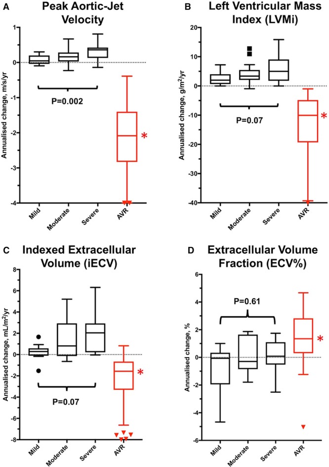
Asymptomatic and symptomatic patients with aortic stenosis underwent repeat echocardiography and magnetic resonance imaging. Changes in peak aortic-jet velocity, left ventricular mass index, diffuse fibrosis (indexed extracellular volume), and replacement fibrosis (late gadolinium enhancement [LGE]) were quantified.
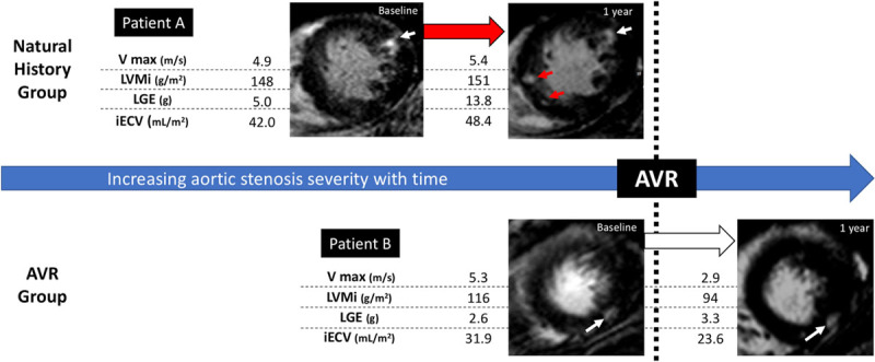
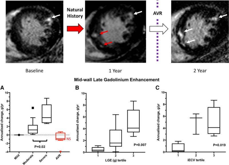
In 61 asymptomatic patients (43% mild, 34% moderate, and 23% severe aortic stenosis), significant increases in peak aortic-jet velocity, left ventricular mass index, indexed extracellular volume, and LGE mass were observed after 2.1±0.7 years, with the most rapid progression observed in patients with most severe stenosis. Patients with baseline midwall LGE (n=16 [26%]; LGE mass, 2.5 g [0.8-4.8 g]) demonstrated particularly rapid increases in scar burden (78% [50%-158%] increase in LGE mass per year). In 38 symptomatic patients (age, 66±8 years; 76% men) who underwent AVR, there was a 19% (11%-25%) reduction in left ventricular mass index (<0.0001) and an 11% (4%-16%) reduction in indexed extracellular volume (=0.003) 0.9±0.3 years after surgery. By contrast midwall LGE (n=10 [26%]; mass, 3.3 g [2.6-8.0 g]) did not change post-AVR (n=10; 3.5 g [2.1-8.0 g]; =0.23), with no evidence of regression even out to 2 years.
In patients with aortic stenosis, cellular hypertrophy and diffuse fibrosis progress in a rapid and balanced manner but are reversible after AVR. Once established, midwall LGE also accumulates rapidly but is irreversible post valve replacement. Given its adverse long-term prognosis, prompt AVR when midwall LGE is first identified may improve clinical outcomes.
URL: https://www.clinicaltrials.gov. Unique identifiers: NCT01755936 and NCT01679431.
主动脉瓣狭窄伴有进行性左心室肥厚和纤维化。我们研究了无症状患者这些过程的自然史及其主动脉瓣置换术后(AVR)的潜在逆转。
无症状和有症状的主动脉瓣狭窄患者接受重复超声心动图和磁共振成像检查。定量测量峰值主动脉射流速度、左心室质量指数、弥漫性纤维化(细胞外容积指数)和替代性纤维化(晚期钆增强[LGE])的变化。
在 61 名无症状患者(43%为轻度,34%为中度,23%为重度主动脉瓣狭窄)中,经过 2.1±0.7 年,观察到峰值主动脉射流速度、左心室质量指数、细胞外容积指数和 LGE 质量显著增加,在最严重狭窄的患者中观察到最快的进展。基线中层壁 LGE(n=16 [26%];LGE 质量,2.5g [0.8-4.8g])患者的瘢痕负荷增加特别快(每年 LGE 质量增加 78%[50%-158%])。在 38 名接受 AVR 的有症状患者(年龄,66±8 岁;76%为男性)中,左心室质量指数降低 19%(11%-25%)(<0.0001),细胞外容积指数降低 11%(4%-16%)(=0.003),术后 0.9±0.3 年。相比之下,中层壁 LGE(n=10 [26%];质量,3.3g [2.6-8.0g])在 AVR 后没有变化(n=10;3.5g [2.1-8.0g];=0.23),甚至在 2 年时也没有出现逆转的证据。
在主动脉瓣狭窄患者中,细胞肥大和弥漫性纤维化以快速和平衡的方式进展,但在 AVR 后是可逆的。一旦建立,中层壁 LGE 也会迅速积累,但在瓣膜置换后是不可逆转的。鉴于其不良的长期预后,当首次发现中层壁 LGE 时,及时进行 AVR 可能会改善临床结果。
网址:https://www.clinicaltrials.gov。唯一标识符:NCT01755936 和 NCT01679431。