Cell Death and Aging Team, Gustave Roussy Cancer Campus, 114 rue Edouard Vaillant, F-94805, Villejuif, France.
Laboratory of Molecular Radiotherapy, INSERM U1030, Gustave Roussy Cancer Campus, 114 rue Edouard Vaillant, F-94805, Villejuif, France.
Cell Death Dis. 2018 Jun 18;9(7):716. doi: 10.1038/s41419-018-0747-y.
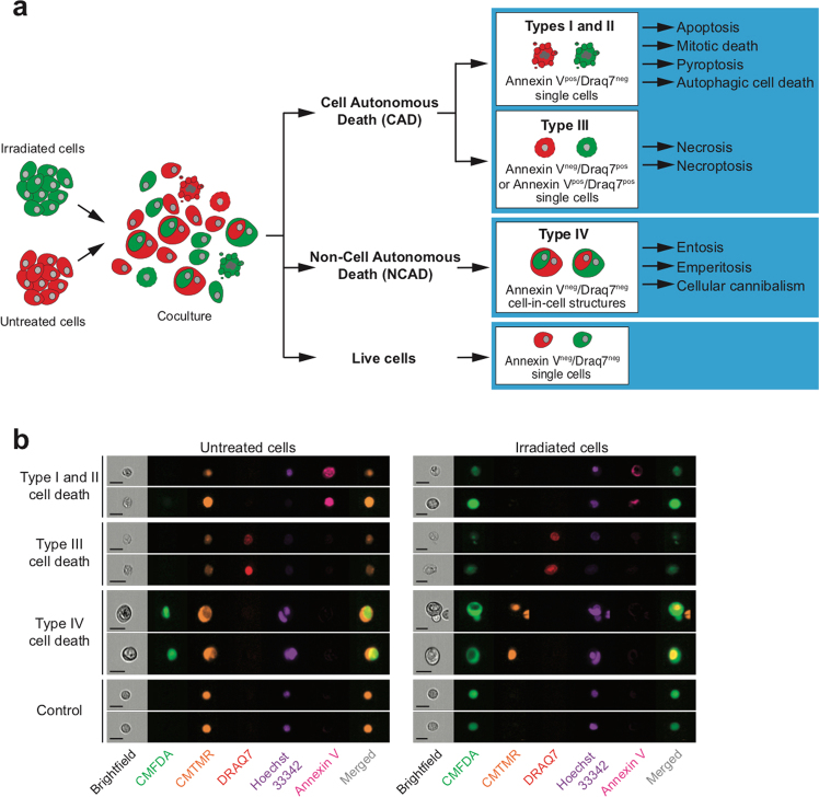
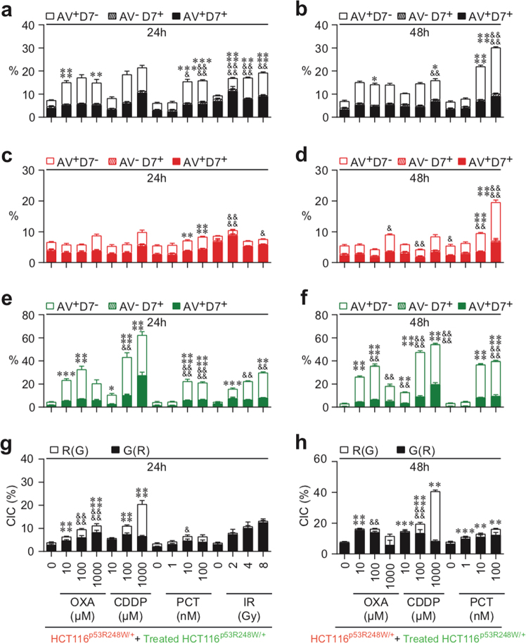
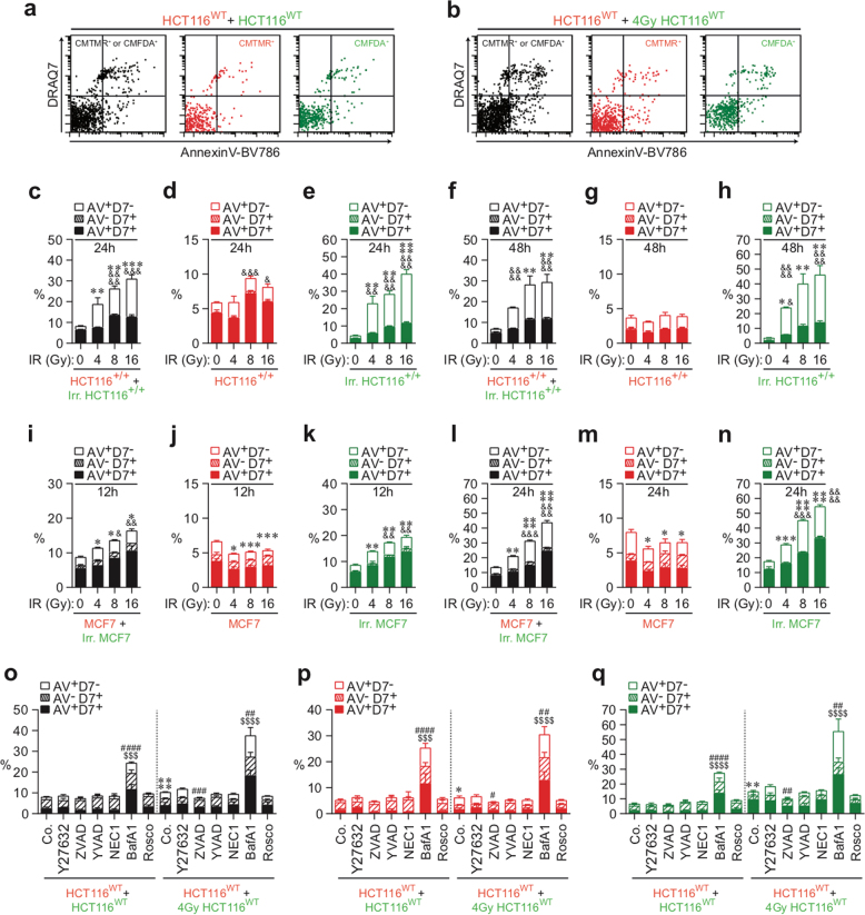
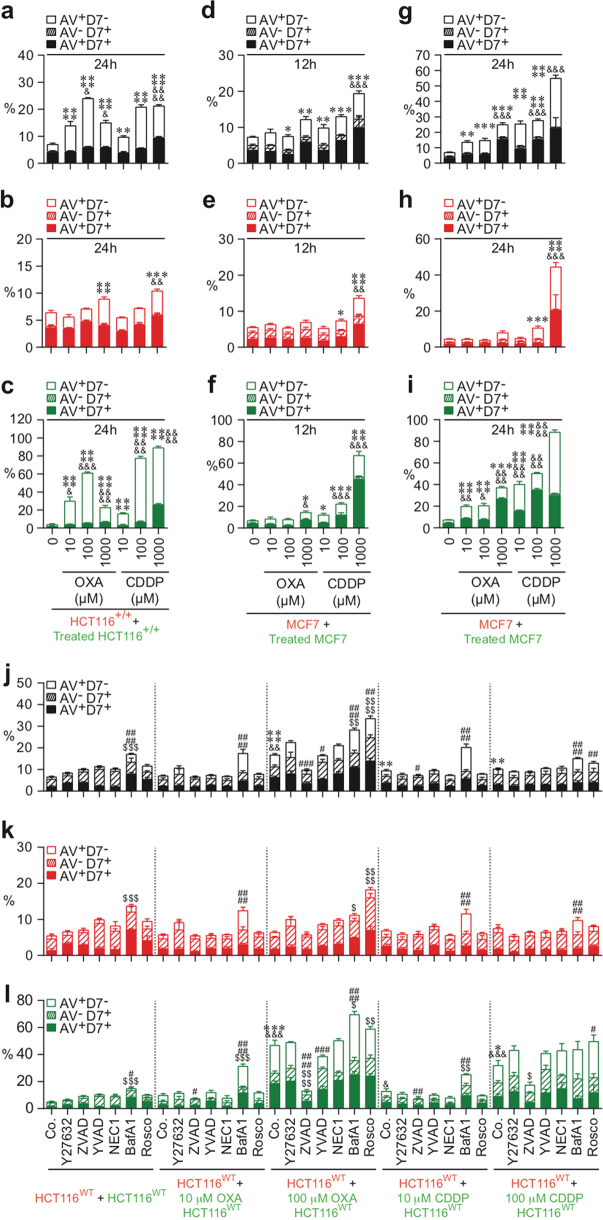
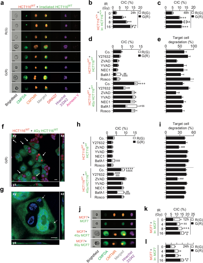
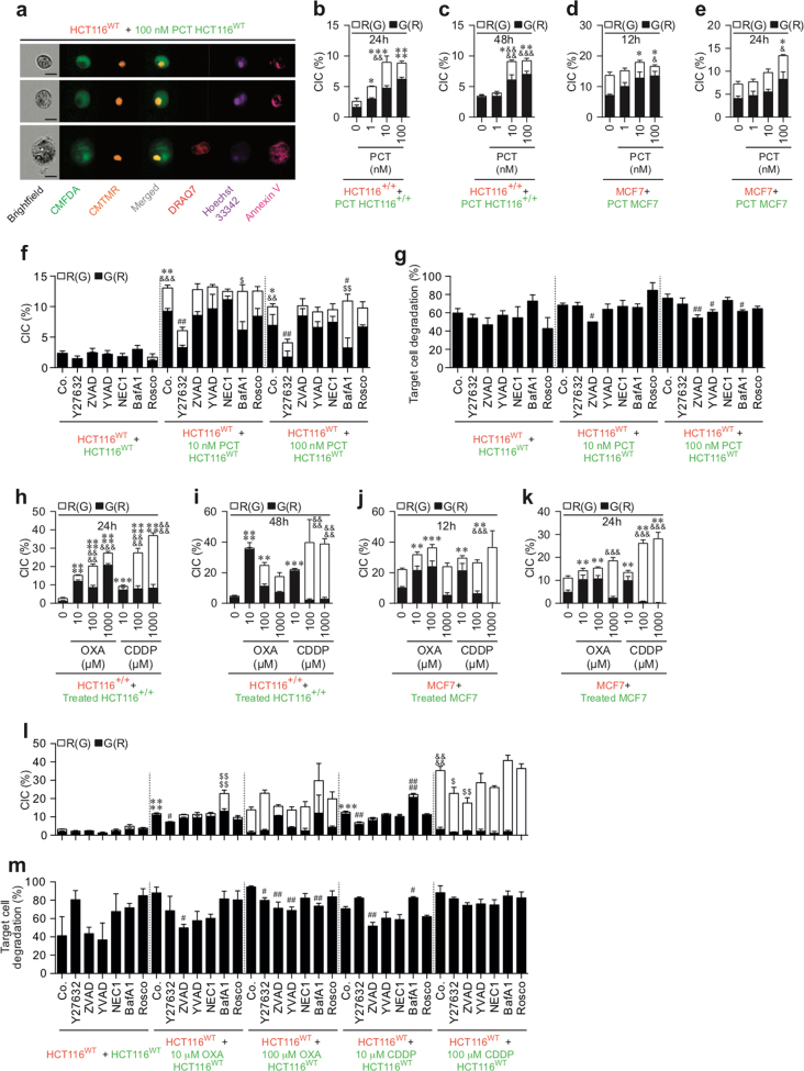
Even though cell death modalities elicited by anticancer chemotherapy and radiotherapy have been extensively studied, the ability of anticancer treatments to induce non-cell-autonomous death has never been investigated. By means of multispectral imaging flow-cytometry-based technology, we analyzed the lethal fate of cancer cells that were treated with conventional anticancer agents and co-cultured with untreated cells, observing that anticancer agents can simultaneously trigger cell-autonomous and non-cell-autonomous death in treated and untreated cells. After ionizing radiation, oxaliplatin, or cisplatin treatment, fractions of treated cancer cell populations were eliminated through cell-autonomous death mechanisms, while other fractions of the treated cancer cells engulfed and killed neighboring cells through non-cell-autonomous processes, including cellular cannibalism. Under conditions of treatment with paclitaxel, non-cell-autonomous and cell-autonomous death were both detected in the treated cell population, while untreated neighboring cells exhibited features of apoptotic demise. The transcriptional activity of p53 tumor-suppressor protein contributed to the execution of cell-autonomous death, yet failed to affect the non-cell-autonomous death by cannibalism for the majority of tested anticancer agents, indicating that the induction of non-cell-autonomous death can occur under conditions in which cell-autonomous death was impaired. Altogether, these results reveal that chemotherapy and radiotherapy can induce both non-cell-autonomous and cell-autonomous death of cancer cells, highlighting the heterogeneity of cell death responses to anticancer treatments and the unsuspected potential contribution of non-cell-autonomous death to the global effects of anticancer treatment.
尽管抗癌化疗和放疗所引发的细胞死亡模式已被广泛研究,但抗癌治疗诱导非细胞自主死亡的能力从未被研究过。通过基于多光谱成像流式细胞术的技术,我们分析了用常规抗癌药物处理并与未处理细胞共培养的癌细胞的致死命运,观察到抗癌药物可以同时在处理和未处理的细胞中触发细胞自主和非细胞自主死亡。在电离辐射、奥沙利铂或顺铂处理后,经细胞自主死亡机制消除了处理的癌细胞群体的分数,而处理的癌细胞的其他分数通过包括细胞自噬在内的非细胞自主过程吞噬和杀死邻近细胞。在紫杉醇处理条件下,在处理的细胞群体中同时检测到非细胞自主和细胞自主死亡,而未处理的邻近细胞表现出凋亡死亡的特征。p53 肿瘤抑制蛋白的转录活性有助于执行细胞自主死亡,但未能影响大多数测试的抗癌药物的细胞自主死亡的非细胞自主吞噬,表明在细胞自主死亡受损的情况下可以诱导非细胞自主死亡。总之,这些结果表明化疗和放疗可以诱导癌细胞的非细胞自主和细胞自主死亡,突出了细胞对抗癌治疗的死亡反应的异质性,以及非细胞自主死亡对抗癌治疗整体效果的潜在预期贡献。