Kobayashi Hitoshi, Ebisawa Katsumi, Kambe Miki, Kasai Takatoshi, Suga Hidetaka, Nakamura Kae, Narita Yuji, Ogata Aika, Kamei Yuzuru
Department of Plastic and Reconstructive Surgery, Nagoya University Graduate School of Medicine, Nagoya, Japan.
Department of Endocrinology and Diabetes, Nagoya University Graduate School of Medicine, Nagoya, Japan.
Nagoya J Med Sci. 2018 May;80(2):141-153. doi: 10.18999/nagjms.80.2.141.
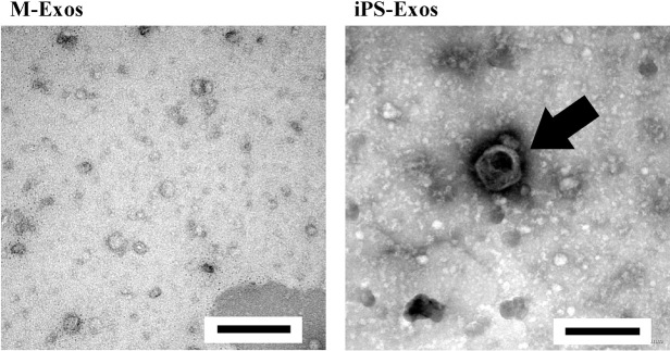
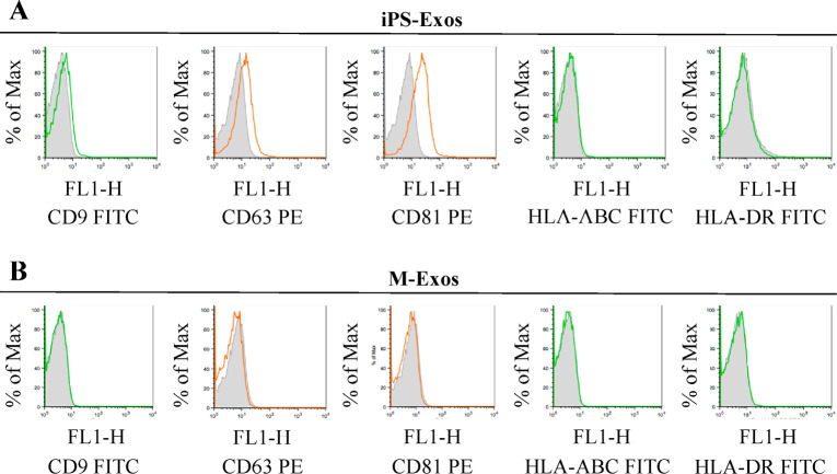
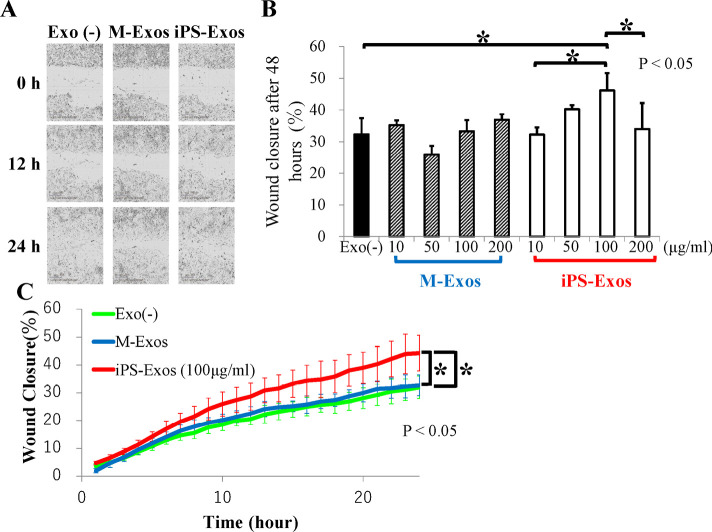
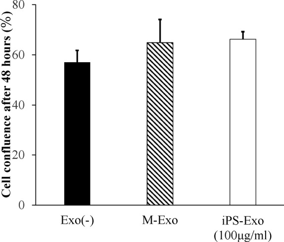
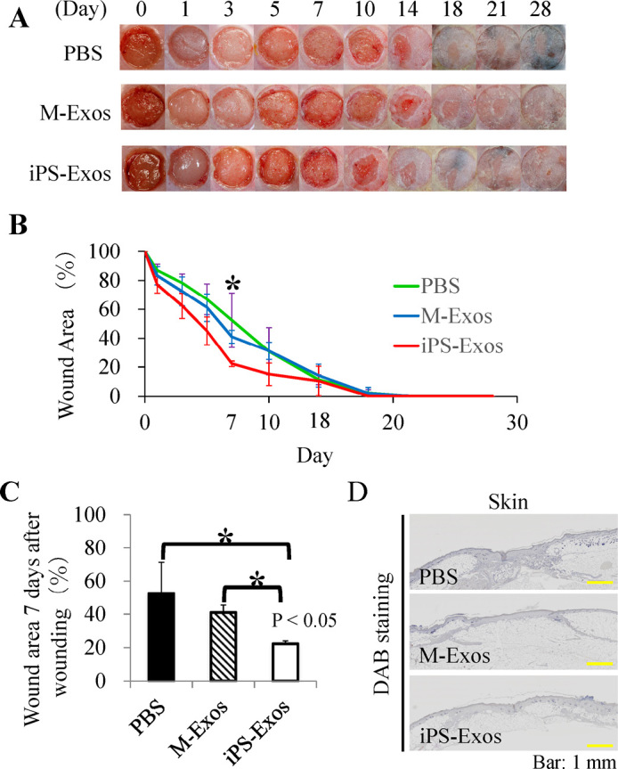
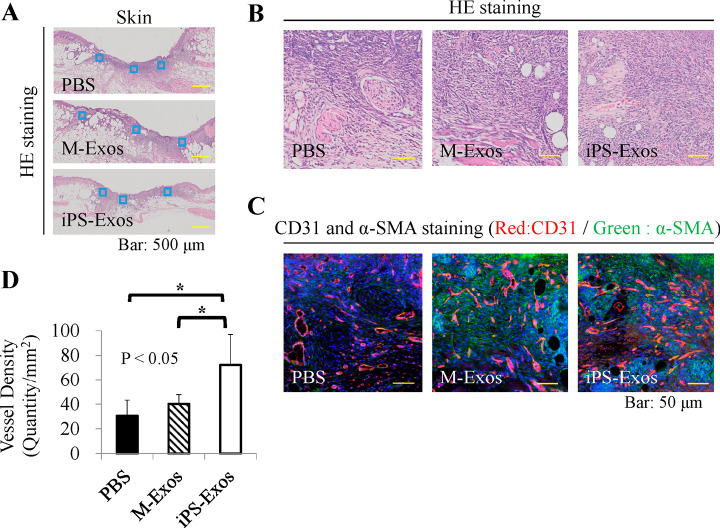
Recently, the effects of stem cell supernatants or exosomes, such as skin wounds, have attracted attention. However, the effects of the induced pluripotent stem (iPS) cell-derived exosomes (iPS-Exos) have not been investigated in detail. Here, we investigated the effects of iPS-Exos on skin wound healing using an animal model. We isolated iPS-Exos from the iPS cell culture media. Control exosomes were isolated from unused iPS cell culture media (M-Exos). We first observed the morphologic characteristics of the isolated exosomes and examined the expression of surface antigens. The effects of these exosomes on the migratory response and proliferation of fibroblasts were analyzed as well. Additionally, using a diabetic ulcer model, the effects of iPS-Exos and M-Exos on skin wound healing were investigated. Transmission electron microscope analysis demonstrated that the size of iPS-Exos (120 ± 25 nm) was significantly larger than that of M-Exos (≤ 100 nm). Flow cytometry analyses showed that iPS-Exos were positive for CD9, CD63, and CD81, whereas they were negative for HLA-ABC and -DR expression. The migratory ability of fibroblasts cocultured with iPS-Exos was shown to be higher than that of the cells cocultured with M-Exos, as demonstrated using scratch assay. Skin wound healing model results showed that the administration of iPS-Exos results in a faster wound closure compared with that observed in the M-Exo group. In conclusion, the results obtained here indicate that iPS-Exos may promote the migration of fibroblasts and , suggesting the possibility of using iPS-Exos for the treatment of diabetic ulcer.
最近,干细胞上清液或外泌体在诸如皮肤伤口等方面的作用引起了关注。然而,诱导多能干细胞(iPS细胞)来源的外泌体(iPS-Exos)的作用尚未得到详细研究。在此,我们使用动物模型研究了iPS-Exos对皮肤伤口愈合的影响。我们从iPS细胞培养基中分离出iPS-Exos。对照外泌体从未使用过的iPS细胞培养基中分离得到(M-Exos)。我们首先观察了分离出的外泌体的形态特征,并检测了表面抗原的表达。还分析了这些外泌体对成纤维细胞迁移反应和增殖的影响。此外,利用糖尿病溃疡模型,研究了iPS-Exos和M-Exos对皮肤伤口愈合的影响。透射电子显微镜分析表明,iPS-Exos的大小(120±25nm)明显大于M-Exos(≤100nm)。流式细胞术分析显示,iPS-Exos对CD9、CD63和CD81呈阳性,而对HLA-ABC和-DR表达呈阴性。划痕试验表明,与iPS-Exos共培养的成纤维细胞的迁移能力高于与M-Exos共培养的细胞。皮肤伤口愈合模型结果显示,与M-Exo组相比,给予iPS-Exos可使伤口愈合更快。总之,此处获得的结果表明,iPS-Exos可能促进成纤维细胞的迁移,提示使用iPS-Exos治疗糖尿病溃疡的可能性。