Department of Orthopaedics, Biomechanics and Implant Technology Research Laboratory, Rostock University Medical Center, Rostock, Germany.
Front Immunol. 2018 Apr 25;9:831. doi: 10.3389/fimmu.2018.00831. eCollection 2018.
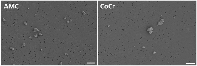
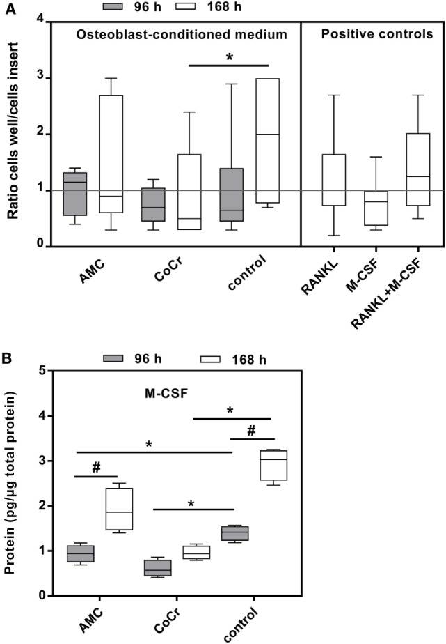
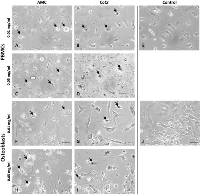
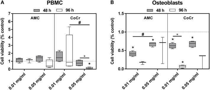
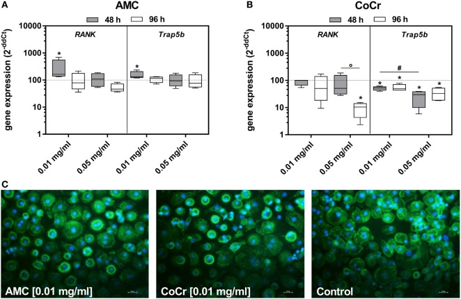
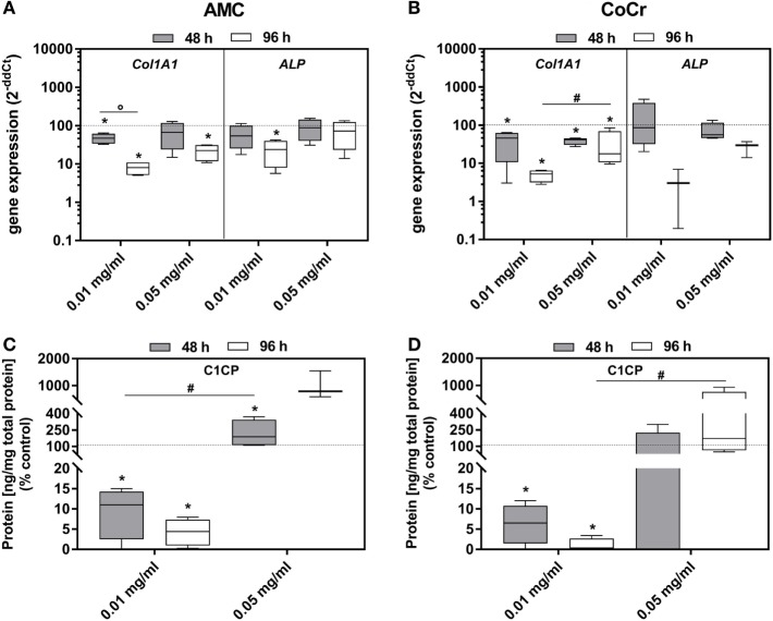
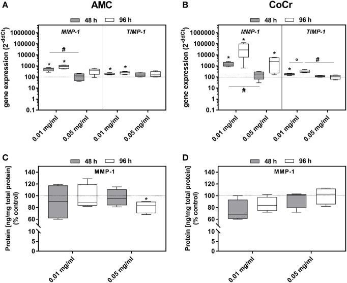
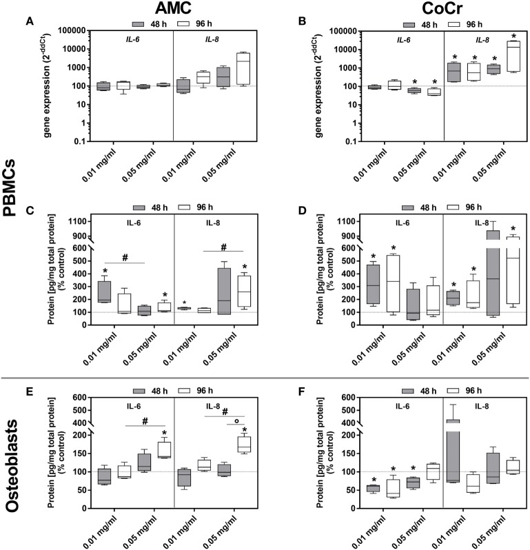
Inflammatory reactions associated with osteolysis and aseptic loosening are the result of wear particles generated at the articulating surfaces of implant components. The aim of the present study was to analyze the biological response of human osteoblasts and peripheral blood mononuclear cells (PBMCs) after exposure to metallic and alumina ceramic particles regarding cellular differentiation, cytokine release, and monocyte migration. Cells were exposed to particles (0.01 and 0.05 mg/ml) from an alumina matrix composite (AMC) ceramic and a CoCr28Mo6 alloy with an average size of 0.5 µm over 48 and 96 h. The expression rates of osteogenic () and pro-osteoclastic () differentiation markers as well as pro-osteolytic mediators () were determined and soluble protein concentrations of active MMP-1, IL-6, IL-8, and pro-collagen type 1 in cell culture supernatants were evaluated. Additionally, the capacity of particle-treated osteoblasts to attract potentially pro-inflammatory cells to the site of particle exposure was investigated by migration assays using osteoblast-conditioned media. The cellular morphology and metabolism of human osteoblasts and adherent PBMCs were influenced by particle type and concentration. In human osteoblasts, expression rates and protein production were significantly reduced after exposing cells to the lower concentration of cobalt-chromium (CoCr) and AMC particles. Exposure to AMC particles (0.01 mg/ml) resulted in increased mRNA levels of and in adherent PBMCs. For gene expression, elevated levels were more prominent after incubation with CoCr compared to AMC particles in osteoblasts, which was not reflected by the protein data. Interleukin (IL)-6 and IL-8 mRNA and protein were induced in both cell types after treatment with AMC particles, whereas exposure to CoCr particles resulted in significantly upregulated IL-6 and IL-8 protein contents in PBMCs only. Exposure of osteoblasts to CoCr particles reduced the chemoattractant potential of osteoblast-conditioned medium. Our results demonstrate distinct effects of AMC and CoCr particles in human osteoblasts and PBMCs. Complex cell and animal models are required to further evaluate the impact of cellular interactions between different cell types during particle exposure.
与骨溶解和无菌性松动相关的炎症反应是植入物组件关节表面产生的磨损颗粒的结果。本研究的目的是分析暴露于金属和氧化铝陶瓷颗粒后,人成骨细胞和外周血单核细胞(PBMC)的细胞分化、细胞因子释放和单核细胞迁移的生物学反应。将细胞暴露于 0.5µm 平均粒径的氧化铝基质复合材料(AMC)陶瓷和 CoCr28Mo6 合金的颗粒(0.01 和 0.05mg/ml)中,分别孵育 48 和 96 小时。测定成骨()和破骨前()分化标志物的表达率以及促溶骨介质()的表达率,并评估细胞培养上清液中活性 MMP-1、IL-6、IL-8 和前胶原 type 1 的可溶性蛋白浓度。此外,通过使用成骨细胞条件培养基进行的迁移实验,研究了受颗粒处理的成骨细胞吸引潜在促炎细胞到颗粒暴露部位的能力。细胞类型和浓度影响人成骨细胞和贴壁 PBMC 的细胞形态和代谢。在人成骨细胞中,当细胞暴露于较低浓度的钴铬(CoCr)和 AMC 颗粒时, 表达率和蛋白产生明显降低。暴露于 AMC 颗粒(0.01mg/ml)导致贴壁 PBMC 中 和 的 mRNA 水平升高。对于 基因表达,与 AMC 颗粒相比,CoCr 颗粒孵育后成骨细胞中的上调水平更为明显,但蛋白数据并未反映这一点。两种细胞类型在用 AMC 颗粒处理后均诱导白细胞介素(IL)-6 和 IL-8 的 mRNA 和蛋白表达,而在用 CoCr 颗粒处理后仅 PBMC 中的 IL-6 和 IL-8 蛋白含量显著上调。成骨细胞暴露于 CoCr 颗粒降低了成骨细胞条件培养基的趋化吸引潜力。我们的结果表明,AMC 和 CoCr 颗粒对人成骨细胞和 PBMC 具有不同的作用。需要复杂的细胞和动物模型来进一步评估颗粒暴露过程中不同细胞类型之间细胞相互作用的影响。