Barbier L, Ramos E, Mendiola J, Rodriguez O, Santamaria G, Santamaria J, Arteagoitia I
University of Basque Country, Departamento de Estomatologia, c/ Barrio Sarriena s/n, 48940 Leioa, Bizkaia,
Med Oral Patol Oral Cir Bucal. 2018 Jul 1;23(4):e469-e477. doi: 10.4317/medoral.22466.
Since the discovery of adult mesenchymal stem cells extensive research has been conducted to determine their mechanisms of differentiation and effectiveness in cell therapy and regenerative medicine.
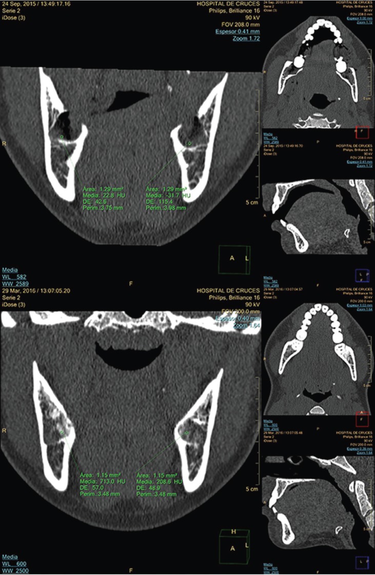
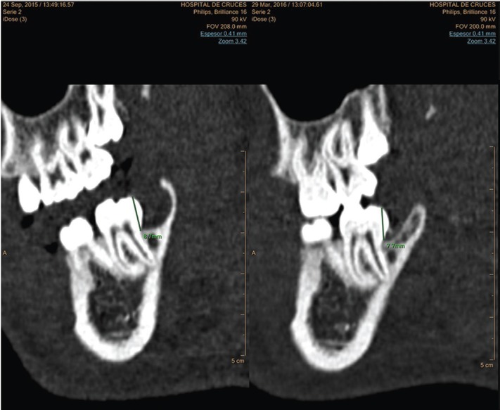
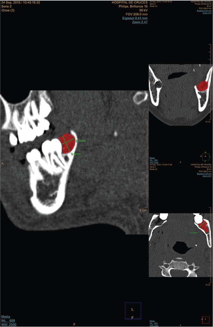
To assess the efficacy of autologous dental pulp mesenchymal stem cells delivered in a collagen matrix for post-extraction socket healing, a single-centre, double-blind, randomised, split-mouth, controlled clinical trial was performed. Both impacted mandibular third molars were extracted from 32 patients. Dental pulp was collected and dissociated; the resulting cell suspension, obtained by centrifugation, was incorporated into a resorbable collagen matrix and implanted in 32 experimental post-extraction sockets. Collagen matrices alone were implanted in 32 contralateral, control post-extraction sockets. Two neuroradiologists independently assessed the extent of bone repair at 6 months after the extractions. Computed tomography (CT, Philips Brilliance) and an advanced display platform (IntelliSpace Portal) was used to record extraction socket density, expressed as Hounsfield units (HU) and height (mm) of the distal interdental bone septum of the second molar. Measurements at 6 months post-extraction were compared with measurements obtained immediately after extraction. Data were analysed with the statistical program STATA 14.
Two patients dropped out of the study. The final sample consisted of 22 women and 8 men (mean age, 23 years; range: 18-30 years). Clinical, radiological, and surgical characteristics of impacted third molars of the control and experimental groups were homogeneous. Measurements obtained by the two neuroradiologists showed agreement. No significant differences were found in the extent of bone repair during analyses of density (p=0.4203 neuroradiologist 1; p=0.2525 neuroradiologist 2) or interdental septum height (p=0.2280 neuroradiologist 1; p=0.4784 neuroradiologist 2).
In our clinical trial, we were unable to demonstrate that autologous dental pulp mesenchymal stem cells reduce socket bone resorption after inferior third molar extraction.
自成人间充质干细胞被发现以来,人们进行了广泛研究,以确定其分化机制以及在细胞治疗和再生医学中的有效性。
为评估在胶原基质中递送的自体牙髓间充质干细胞对拔牙后牙槽窝愈合的疗效,开展了一项单中心、双盲、随机、分口对照临床试验。从32例患者中拔除双侧下颌阻生第三磨牙。收集牙髓并进行解离;通过离心得到的细胞悬液被掺入可吸收胶原基质中,并植入32个拔牙后的试验性牙槽窝。仅将胶原基质植入32个对侧对照拔牙后牙槽窝。两名神经放射科医生在拔牙后6个月独立评估骨修复程度。使用计算机断层扫描(CT,飞利浦Brilliance)和先进的显示平台(IntelliSpace Portal)记录拔牙窝密度,以亨氏单位(HU)表示,以及第二磨牙远中牙间骨间隔的高度(mm)。将拔牙后6个月的测量结果与拔牙后立即获得的测量结果进行比较。使用统计软件STATA 14对数据进行分析。
两名患者退出研究。最终样本包括22名女性和8名男性(平均年龄23岁;范围:18 - 30岁)。对照组和试验组阻生第三磨牙的临床、放射学和手术特征相似。两名神经放射科医生的测量结果一致。在密度分析(神经放射科医生1,p = 0.4203;神经放射科医生2,p = 0.2525)或牙间间隔高度分析(神经放射科医生1,p = 0.2280;神经放射科医生2,p = 0.4784)中,未发现骨修复程度有显著差异。
在我们的临床试验中,未能证明自体牙髓间充质干细胞可减少下颌第三磨牙拔除后牙槽骨吸收。