Fan Wen-Juan, Yan Ming-Chao, Wang Lai, Sun Yi-Zheng, Deng Jin-Bo, Deng Jie-Xin
Institute of Neurobiology, School of Life Science, Henan University, Kaifeng, Henan Province, China.
Neural Regen Res. 2018 Jun;13(6):1019-1025. doi: 10.4103/1673-5374.233445.
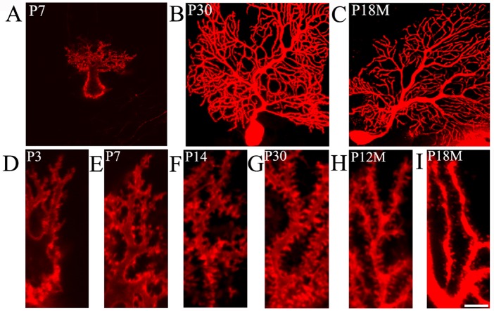
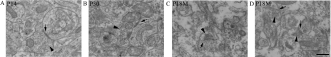
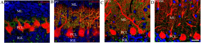
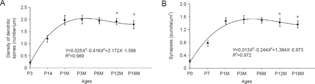
Synapses are key structures in neural networks, and are involved in learning and memory in the central nervous system. Investigating synaptogenesis and synaptic aging is important in understanding neural development and neural degeneration in diseases such as Alzheimer disease and Parkinson's disease. Our previous study found that synaptogenesis and synaptic maturation were harmonized with brain development and maturation. However, synaptic damage and loss in the aging cerebellum are not well understood. This study was designed to investigate the occurrence of synaptic aging in the cerebellum by observing the ultrastructural changes of dendritic spines and synapses in cerebellar Purkinje cells of aging mice. Immunocytochemistry, DiI diolistic assays, and transmission electron microscopy were used to visualize the morphological characteristics of synaptic buttons, dendritic spines and synapses of Purkinje cells in mice at various ages. With synaptic aging in the cerebellum, dendritic spines and synaptic buttons were lost, and the synaptic ultrastructure was altered, including a reduction in the number of synaptic vesicles and mitochondria in presynaptic termini and smaller thin specialized zones in pre- and post-synaptic membranes. These findings confirm that synaptic morphology and function is disrupted in aging synapses, which may be an important pathological cause of neurodegenerative diseases.
突触是神经网络中的关键结构,参与中枢神经系统的学习和记忆。研究突触发生和突触老化对于理解诸如阿尔茨海默病和帕金森病等疾病中的神经发育和神经退行性变至关重要。我们之前的研究发现,突触发生和突触成熟与大脑发育和成熟是协调一致的。然而,衰老小脑中的突触损伤和丧失情况尚未得到充分了解。本研究旨在通过观察衰老小鼠小脑浦肯野细胞中树突棘和突触的超微结构变化,来研究小脑中突触老化的发生情况。采用免疫细胞化学、DiI 示踪分析和透射电子显微镜来观察不同年龄段小鼠浦肯野细胞的突触小结、树突棘和突触的形态特征。随着小脑突触老化,树突棘和突触小结丧失,突触超微结构发生改变,包括突触前终末中突触小泡和线粒体数量减少,以及突触前膜和突触后膜中较小的薄特化区域。这些发现证实,衰老突触中的突触形态和功能受到破坏,这可能是神经退行性疾病的一个重要病理原因。