Huang Lichao, Zhang Zihao, Qu Baolin, Cui Zhiqiang, Wang Yao, Li Jiwei, Wang Jinyuan, Zuo Zhentao, Zhuo Yan, Yu Xinguang, Lin Zhipei, Pan Longsheng
Department of Neurosurgery, PLA General Hospital, Beijing, CHN.
State Key Laboratory of Brain and Cognitive Science, Institute of Biophysics, Chinese Academy of Sciences.
Cureus. 2018 Apr 18;10(4):e2502. doi: 10.7759/cureus.2502.
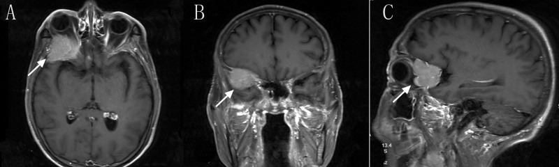
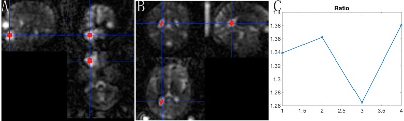
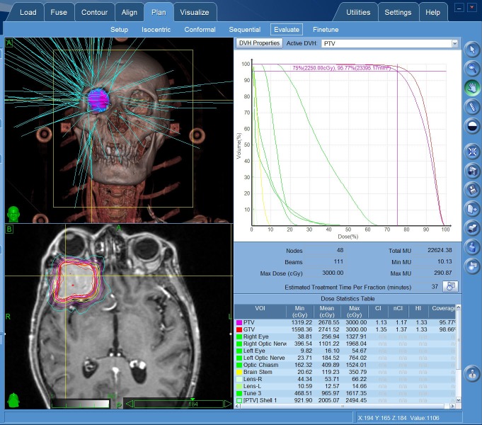
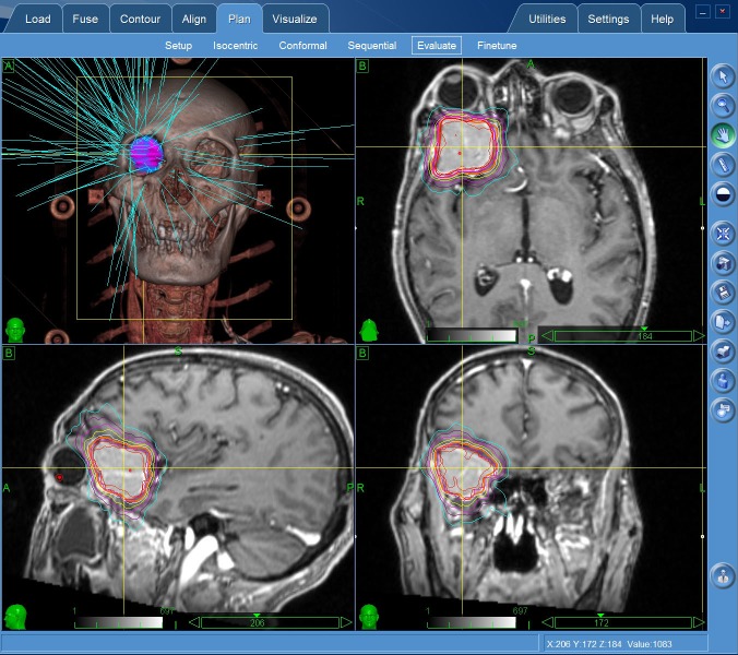
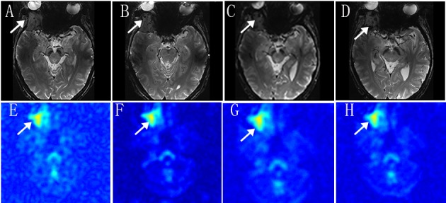
Herein we describe the case of an elderly patient who presented with a recent history of impaired vision of the right eye around three months due to brain lesions. He was diagnosed with liver cancer and underwent surgery three months prior. The pathological result is hepatocellular carcinoma. Magnetic resonance imaging (MRI) revealed the diagnosis of brain to be metastatic. The patient selected CyberKnife (Accuray Incorporated, Sunnyvale, USA) radiosurgery for the brain lesion since his physical conditions are not suitable for craniotomy. We adapt the imaging of sodium MRI and proton diffusion mapping at 7T MR system to evaluate the efficacy following CyberKnife early stage treatment. To date, we find the tissue sodium concentration (TSC) changes with the time whereas the proton MRI has no significant change within one month. The time course of sodium concentration in the tumor showed a dramatic increase in the treated brain tumor compared to the pretreatment sodium concentration and 48 hours after stereotactic radiosurgery (SRS), which is correlated to the period of the radiotherapy-induced cellular necrosis. This case demonstrates the possibility of sodium MRI as a biomarker for monitoring early radiotherapy for assessing tumor cellularity.
在此,我们描述了一位老年患者的病例,该患者因脑病变出现右眼视力受损近三个月。他三个月前被诊断为肝癌并接受了手术。病理结果为肝细胞癌。磁共振成像(MRI)显示脑部诊断为转移性病变。由于患者身体状况不适合开颅手术,故选择射波刀(美国加利福尼亚州森尼韦尔市Accuray公司)对脑部病变进行放射外科治疗。我们采用7T MR系统的钠MRI成像和质子扩散成像来评估射波刀早期治疗后的疗效。迄今为止,我们发现组织钠浓度(TSC)随时间变化,而质子MRI在一个月内无显著变化。与立体定向放射外科(SRS)治疗前及治疗后48小时的钠浓度相比,肿瘤内钠浓度的时间进程显示治疗后的脑肿瘤显著增加,这与放疗诱导的细胞坏死期相关。该病例证明了钠MRI作为监测早期放疗以评估肿瘤细胞密度生物标志物的可能性。