Woo Taylor E, Lim Rachel, Surette Michael G, Waddell Barbara, Bowron Joel C, Somayaji Ranjani, Duong Jessica, Mody Christopher H, Rabin Harvey R, Storey Douglas G, Parkins Michael D
Dept of Biological Sciences, University of Calgary, Calgary, Canada.
Dept of Medicine, University of Calgary, Calgary, Canada.
ERJ Open Res. 2018 Jun 18;4(2). doi: 10.1183/23120541.00162-2017. eCollection 2018 Apr.
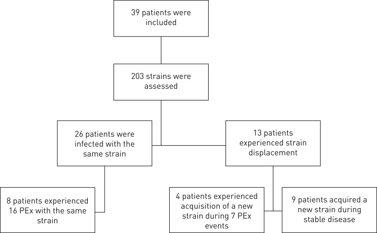
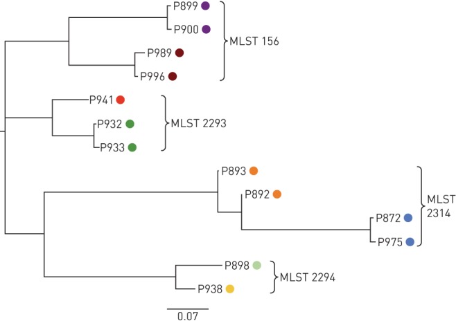
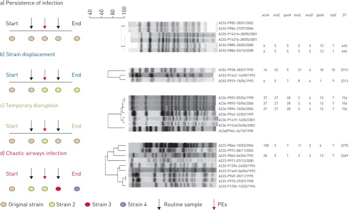
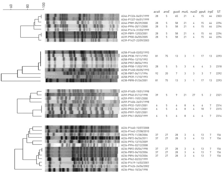
The natural history and epidemiology of infections in non-cystic fibrosis (non-CF) bronchiectasis is not well understood. As such it was our intention to determine the evolution of airway infection and the transmission potential of in patients with non-CF bronchiectasis. A longitudinal cohort study was conducted from 1986-2011 using a biobank of prospectively collected isolates from patients with non-CF bronchiectasis. Patients included were ≥18 years old and had ≥2 positive cultures over a minimum 6-month period. All isolates obtained at first and most recent clinical encounters, as well as during exacerbations, that were morphologically distinct on MacConkey agar were genotyped by pulsed-field gel electrophoresis (PFGE) and multilocus sequence typing (MLST). A total of 203 isolates from 39 patients were analysed. These were compared to a large collection of globally epidemic and local CF strains, as well as non-CF isolates. We identified four patterns of infection in non-CF bronchiectasis including: 1) persistence of a single strain (n=26; 67%); 2) strain displacement (n=8; 20%); 3) temporary disruption (n=3; 8%); and 4) chaotic airway infection (n=2; 5%). Patterns of infection were not significant predictors of rates of lung function decline or progression to end-stage disease and acquisition of new strains did not associate with the occurrence of exacerbations. Rarely, non-CF bronchiectasis strains with similar pulsotypes were observed in CF and non-CF controls, but no CF epidemic strains were observed. While rare shared strains were observed in non-CF bronchiectasis, whole-genome sequencing refuted patient-patient transmission. We observed a higher incidence of strain-displacement in our patient cohort compared to those observed in CF studies, although this did not impact on outcomes.
非囊性纤维化(非CF)支气管扩张症感染的自然史和流行病学尚未得到充分了解。因此,我们旨在确定非CF支气管扩张症患者气道感染的演变情况及其传播潜力。我们于1986年至2011年开展了一项纵向队列研究,使用了一个生物样本库,该样本库前瞻性收集了非CF支气管扩张症患者的分离株。纳入的患者年龄≥18岁,在至少6个月的时间内有≥2次阳性培养结果。在首次和最近一次临床就诊时以及病情加重期间获得的所有在麦康凯琼脂上形态不同的分离株,通过脉冲场凝胶电泳(PFGE)和多位点序列分型(MLST)进行基因分型。共分析了39例患者的203株分离株。将这些分离株与大量全球流行和本地CF菌株以及非CF分离株进行了比较。我们在非CF支气管扩张症中确定了四种感染模式,包括:1)单一菌株持续存在(n = 26;67%);2)菌株替代(n = 8;20%);3)暂时中断(n = 3;8%);4)气道感染混乱(n = 2;5%)。感染模式并非肺功能下降率或进展至终末期疾病的显著预测因素,新菌株的获得与病情加重的发生无关。在CF和非CF对照中很少观察到具有相似脉冲型的非CF支气管扩张症菌株,但未观察到CF流行菌株。虽然在非CF支气管扩张症中观察到了罕见的共享菌株,但全基因组测序排除了患者之间的传播。与CF研究中观察到的情况相比,我们的患者队列中菌株替代的发生率更高,尽管这并未影响结果。