Joint Department of Biomedical Engineering, University of North Carolina, Chapel Hill, and North Carolina State University, Raleigh, North Carolina, USA.
Lab Chip. 2018 Jul 24;18(15):2202-2213. doi: 10.1039/c8lc00332g.
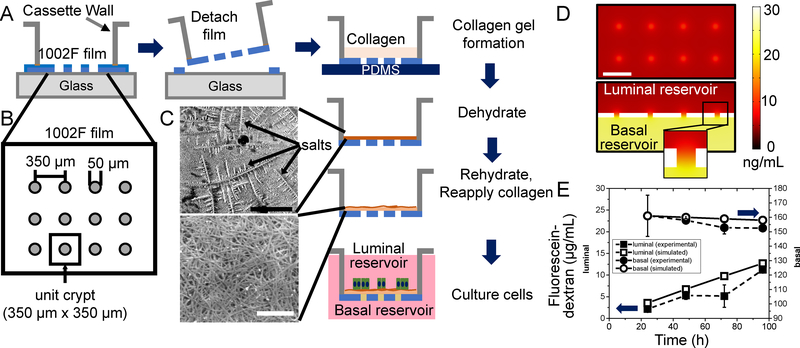
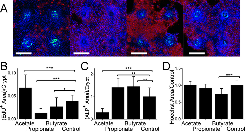
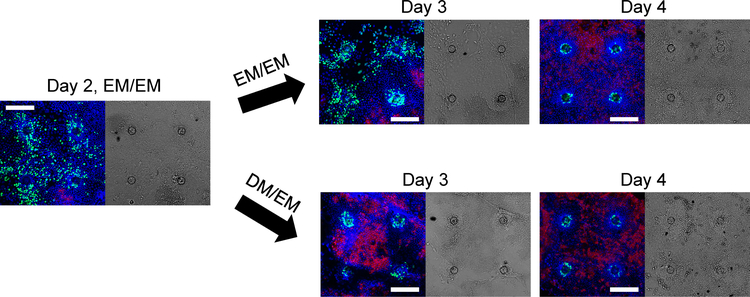
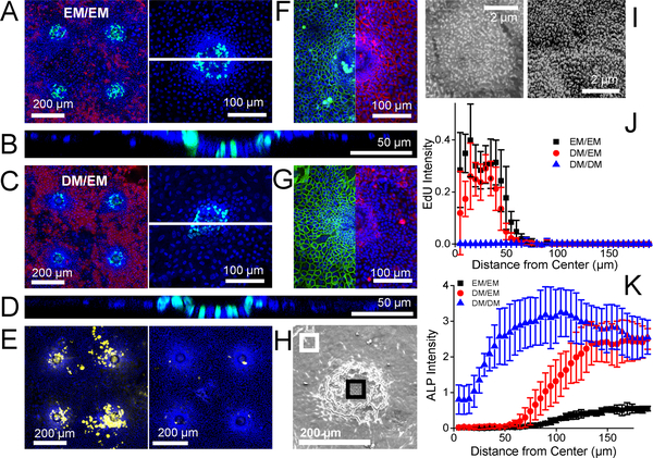
A simple, in vitro intestinal model recapitulating key aspects of crypt architecture and physiology would facilitate our understanding the impact of drugs, foods and microbial metabolites on the intestine. To address the limitations of previously reported intestinal in vitro platforms, we developed a planar crypt array that replicated the spatial segregation and physiologic responses of primary mouse intestinal epithelial cells in the large intestine. Collagen was coated across an impermeable film possessing an array of microholes creating two regions of distinct stiffness and porosity (above and outside the microholes). Primary mouse colon epithelial cells formed a continuous monolayer across the array with a proliferative cell zone above the microholes and a nonproliferative or differentiated cell region distant from the microholes. Formation of a chemical gradient of growth factors across the array yielded a more complete or in vivo-like cell segregation of proliferative and differentiated cells with cell migration outward from the proliferative cell zone into the differentiated zone to replace apoptotic dying cells much as occurs in vivo. Short chain fatty acids (microbial metabolites) applied to the luminal surface of the crypt array significantly impacted the proliferation and differentiation of the cells replicating the known in vivo effects of these fatty acids. Importantly this planar crypt array was readily fabricated and maintained, easily imaged with properties quantified by microscopy, and compatible with reagent addition to either the luminal or basal fluid reservoirs. The ability to observe simultaneously stem/proliferative and differentiated cell behavior and movement between these two compartments in response to drugs, toxins, inflammatory mediators or microbial metabolites will be of widespread utility.
一种简单的、体外肠道模型可以重现隐窝结构和生理学的关键方面,有助于我们了解药物、食物和微生物代谢物对肠道的影响。为了解决以前报道的肠道体外平台的局限性,我们开发了一种平面隐窝阵列,复制了主要的小鼠肠道上皮细胞在大肠中的空间分离和生理反应。胶原蛋白涂覆在不可渗透的薄膜上,该薄膜上有一系列微孔,形成两个具有明显硬度和孔隙率的区域(微孔上方和外侧)。原代小鼠结肠上皮细胞在阵列上形成一个连续的单层,微孔上方是有丝分裂细胞区,远离微孔的是无丝分裂或分化细胞区。在阵列上形成生长因子的化学梯度,产生更完整或更类似于体内的增殖和分化细胞的细胞分离,细胞从有丝分裂细胞区向外迁移到分化区,以替代体内发生的凋亡死亡细胞。短链脂肪酸(微生物代谢物)施加到隐窝阵列的腔表面显著影响细胞的增殖和分化,复制了这些脂肪酸的已知体内效应。重要的是,这种平面隐窝阵列易于制造和维护,通过显微镜可以轻松成像,并可以与试剂添加到腔或基底液储器中兼容。观察这些两种隔室之间的干细胞/增殖和分化细胞行为和运动对药物、毒素、炎症介质或微生物代谢物的同时响应的能力将具有广泛的用途。