Department of Diagnostic and Interventional Radiology, Lausanne University Hospital, Rue du Bugnon 46, CH-1011, Lausanne, Switzerland.
Advanced Clinical Imaging Technology, Siemens Healthcare HC CEMEA SUI DI PI, Lausanne, Switzerland.
Brain Imaging Behav. 2019 Jun;13(3):810-818. doi: 10.1007/s11682-018-9909-x.
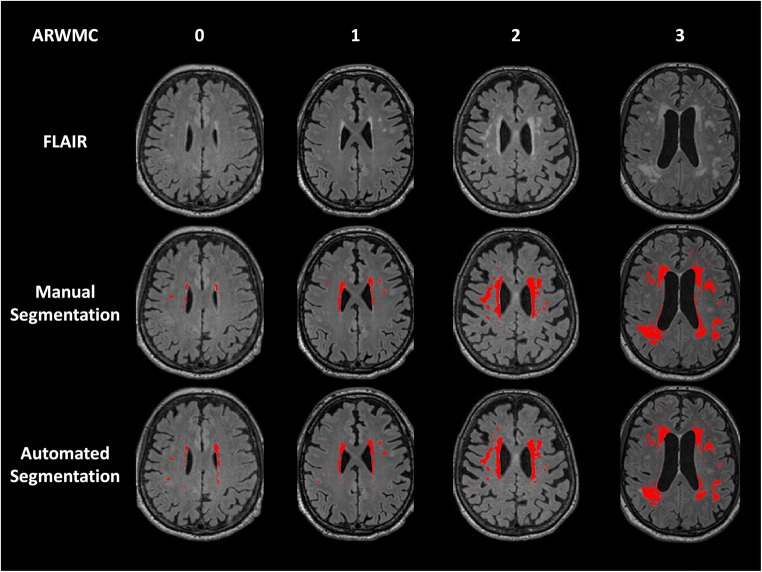
The relation of white matter hyperintense lesions to episodic memory impairment in patients with Parkinson's disease (PD) is still controversial. We aimed at evaluating the relation between white matter hyperintense lesions and episodic memory decline in patients with PD. In this multicentric prospective study, twenty-one normal controls, 15 PD patients without mild cognitive impairment (MCI) and 13 PD patients with MCI were selected to conduct a clinico-radiological correlation analysis. Performance during episodic memory testing, age-related white matter changes score, total manual and automated white matter hyperintense lesions volume and lobar white matter hyperintense lesions volumes were compared between groups using the Kruskal-Wallis and Wilcoxon signed-rank tests, and correlations were assessed using the Spearman test. MCI PD patients had impaired free recall. They also had higher total, left prefrontal and left temporal white matter hyperintense lesions volumes than normal controls. Free recall performance was negatively correlated with the total white matter hyperintense lesions volume, either manually or automatically delineated, but not with the age-related white matter changes score. Using automated segmentation, both the left prefrontal and temporal white matter hyperintense lesions volumes were negatively correlated with the free recall performance. Early episodic memory impairment in MCI PD patients may be related to white matter hyperintense lesions, mainly in the prefrontal and temporal lobes. This relation is influenced by the method used for white matter hyperintense lesions quantification. Automated volumetry allows for detecting those changes.
帕金森病(PD)患者的脑白质高信号(WMH)与情景记忆障碍的关系仍存在争议。我们旨在评估 PD 患者的脑白质高信号与情景记忆下降之间的关系。在这项多中心前瞻性研究中,我们选择了 21 名正常对照、15 名无轻度认知障碍(MCI)的 PD 患者和 13 名 MCI PD 患者,进行临床-放射学相关性分析。采用 Kruskal-Wallis 和 Wilcoxon 符号秩检验比较各组之间的情景记忆测试表现、年龄相关性脑白质改变评分、总手动和自动脑白质高信号病变体积以及脑叶脑白质高信号病变体积,并采用 Spearman 检验评估相关性。MCI PD 患者的自由回忆能力受损。与正常对照组相比,他们的总脑白质高信号病变体积、左额极和左颞叶脑白质高信号病变体积更高。自由回忆表现与总脑白质高信号病变体积(无论是手动还是自动勾画)呈负相关,但与年龄相关性脑白质改变评分无关。使用自动分割,左额极和颞叶脑白质高信号病变体积与自由回忆表现均呈负相关。MCI PD 患者的早期情景记忆障碍可能与脑白质高信号病变有关,主要位于额极和颞叶。这种关系受脑白质高信号病变定量方法的影响。自动容积测量法可检测到这些变化。