Mohajeri Moghaddam Sarah, Bhatt Alok A
Department of Imaging Sciences, University of Rochester Medical Center, 601 Elmwood Avenue, P.O. Box 648, Rochester, NY, 14642, USA.
Insights Imaging. 2018 Aug;9(4):511-526. doi: 10.1007/s13244-018-0608-3. Epub 2018 Jun 12.
Intramedullary spinal cord abnormalities are often challenging to diagnose. Spinal cord biopsy is a high-risk procedure with the potential to cause permanent neurological injury. Magnetic resonance imaging is the modality of choice for diagnosis and preoperative assessment of patients with spinal cord abnormalities. The radiologist's ability to narrow the differential diagnosis of spinal cord abnormalities has the potential to save patients from invasive approaches for diagnosis and also guide appropriate management.
APPROACH/METHODS: This article will provide a systematic approach to the evaluation of intramedullary spinal cord lesions-with emphasis on location, length and segment distribution, and enhancement pattern-to help narrow the differential diagnosis. In doing so, we will review various spinal cord pathologies, including demyelinating and metabolic conditions, neoplasms, and vascular lesions. Although intramedullary spinal cord abnormalities can be a challenge for the radiologist, a systematic approach to the differential diagnosis with a focus on lesion location, cord length and segment involvement, as well as enhancement pattern, can greatly help narrow the differential diagnosis, if not synch the diagnosis. This strategy will potentially obviate the need for an invasive approach to diagnosis and help guide treatment.
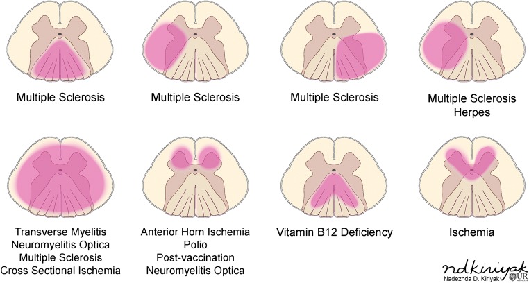
• Imaging diagnosis of intramedullary spinal cord lesions could obviate cord biopsy. • Evaluation of cord lesions should focus on location, length, and enhancement pattern. • In demyelination, the degree of cross-sectional involvement is a distinguishing feature.
脊髓髓内异常的诊断往往具有挑战性。脊髓活检是一种高风险操作,有可能导致永久性神经损伤。磁共振成像(MRI)是诊断脊髓异常患者及进行术前评估的首选方式。放射科医生缩小脊髓异常鉴别诊断范围的能力有可能使患者避免采用侵入性诊断方法,并指导恰当的治疗。
本文将提供一种评估脊髓髓内病变的系统方法,重点关注病变位置、长度及节段分布以及强化方式,以帮助缩小鉴别诊断范围。在此过程中,我们将回顾各种脊髓病变,包括脱髓鞘和代谢性疾病、肿瘤及血管病变。尽管脊髓髓内异常对放射科医生来说可能是一项挑战,但一种以病变位置、脊髓长度及受累节段以及强化方式为重点的系统鉴别诊断方法,即使不能确诊,也能极大地帮助缩小鉴别诊断范围。这一策略有可能避免采用侵入性诊断方法,并有助于指导治疗。
• 脊髓髓内病变的影像学诊断可避免脊髓活检。• 对脊髓病变的评估应关注位置、长度及强化方式。• 在脱髓鞘疾病中,横断面受累程度是一个显著特征。