• 文献检索
  • 文档翻译
  • 深度研究
  • 学术资讯
  • Suppr Zotero 插件Zotero 插件
  • 邀请有礼
  • 套餐&价格
  • 历史记录
应用&插件
Suppr Zotero 插件Zotero 插件浏览器插件Mac 客户端Windows 客户端微信小程序
定价
高级版会员购买积分包购买API积分包
服务
文献检索文档翻译深度研究API 文档MCP 服务
关于我们
关于 Suppr公司介绍联系我们用户协议隐私条款
关注我们

Suppr 超能文献

核心技术专利:CN118964589B侵权必究
粤ICP备2023148730 号-1Suppr @ 2026

文献检索

告别复杂PubMed语法,用中文像聊天一样搜索,搜遍4000万医学文献。AI智能推荐,让科研检索更轻松。

立即免费搜索

文件翻译

保留排版,准确专业,支持PDF/Word/PPT等文件格式,支持 12+语言互译。

免费翻译文档

深度研究

AI帮你快速写综述,25分钟生成高质量综述,智能提取关键信息,辅助科研写作。

立即免费体验

古埃及卡诺匹斯罐的放射学发现:比较三种标准临床成像方式(X射线、CT和MRI)。

Radiological findings in ancient Egyptian canopic jars: comparing three standard clinical imaging modalities (x-rays, CT and MRI).

作者信息

Eppenberger Patrick E, Cavka Mislav, Habicht Michael E, Galassi Francesco M, Rühli Frank

机构信息

1Institute of Evolutionary Medicine, University of Zurich, Winterthurerstrasse 190, CH-8057 Zurich, Switzerland.

2School of Medicine, University of Zagreb, Šalata 3, 10 000 Zagreb, Croatia.

出版信息

Eur Radiol Exp. 2018 Jun 20;2:12. doi: 10.1186/s41747-018-0048-3. eCollection 2018 Dec.

DOI:10.1186/s41747-018-0048-3
PMID:29951641
原文链接:https://pmc.ncbi.nlm.nih.gov/articles/PMC6008346/
Abstract

BACKGROUND

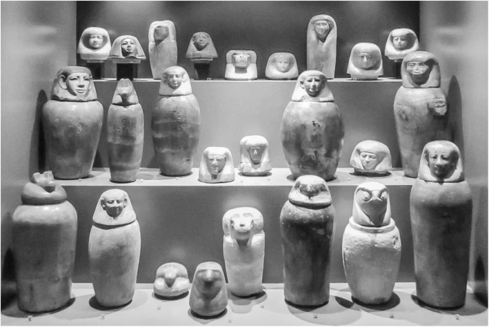
The aim of our study was to evaluate the potential and the limitations of standard clinical imaging modalities for the examination of ancient Egyptian canopic jars and the mummified visceral organs (putatively) contained within them.

METHODS

A series of four ancient Egyptian canopic jars was imaged comparing the three standard clinical imaging modalities: x-rays, computed tomography (CT) and magnetic resonance imaging (MRI). Additionally, imaging-data-based volumetric calculations were performed for quantitative assessment of the jar contents.

RESULTS

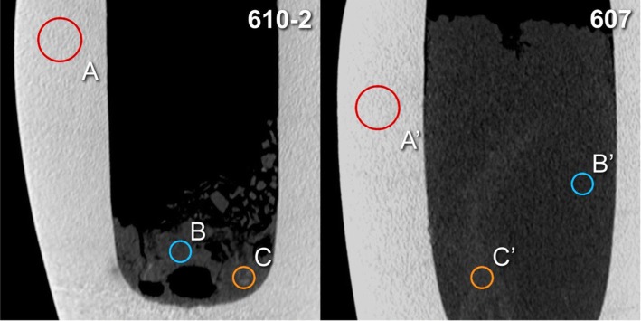
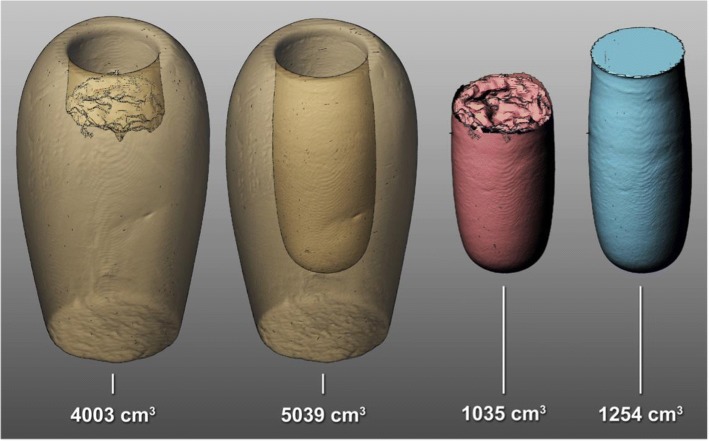
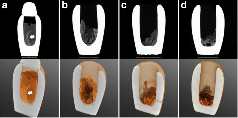
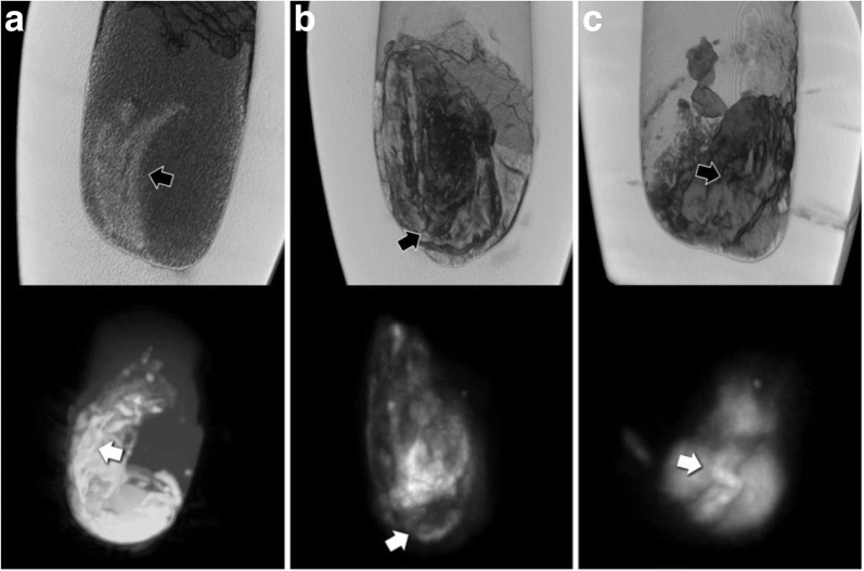
The image contrast of the x-ray images was limited by the thickness and high density of the calcite mineral constituting the examined jars. CT scans showed few artefacts and revealed hyperdense structures of organ-specific morphology, surrounded by a hypodense homogeneous material. The image quality of MRI scans was limited by the low amount of water present in the desiccated jar contents. Nevertheless, areas of pronounced signal intensity coincided well with hyperdense structures previously identified on CT scans. CT-based volumetric calculations revealed holding capacities of the jars of 626-1319 cm and content volumes of 206-1035 cm.

CONCLUSIONS

CT is the modality of choice for non-invasive examination of ancient Egyptian canopic jars. However, despite its limitations, x-ray imaging will often remain the only practicable method for on-site investigations. Overall, the presented radiological findings are more compatible with contained small organ fragments rather than entire mummified organs, as originally expected, with consequent implications for envisioned future sampling for chemical and genetic analysis.

摘要

背景

我们研究的目的是评估标准临床成像方式在检查古埃及石棺罐以及其中(据推测)所含木乃伊化内脏器官方面的潜力和局限性。

方法

对一系列四个古埃及石棺罐进行成像,比较三种标准临床成像方式:x光、计算机断层扫描(CT)和磁共振成像(MRI)。此外,基于成像数据进行体积计算,以对罐内物品进行定量评估。

结果

x光图像的图像对比度受构成被检查罐子的方解石矿物的厚度和高密度限制。CT扫描显示伪影较少,并揭示了具有器官特异性形态的高密度结构,周围是低密度均匀物质。MRI扫描的图像质量受干燥罐内物品中少量水分的限制。然而,明显信号强度区域与先前在CT扫描中识别出的高密度结构吻合良好。基于CT的体积计算显示罐子的容纳容量为626 - 1319立方厘米,内容物体积为206 - 1035立方厘米。

结论

CT是古埃及石棺罐非侵入性检查的首选方式。然而,尽管有其局限性,x光成像通常仍将是现场调查的唯一可行方法。总体而言,所呈现的放射学发现与所含小器官碎片而非最初预期的完整木乃伊化器官更相符,这对未来设想的化学和基因分析采样有相应影响。

https://cdn.ncbi.nlm.nih.gov/pmc/blobs/cd4c/6091675/0f45906266d8/41747_2018_48_Fig6_HTML.jpg
https://cdn.ncbi.nlm.nih.gov/pmc/blobs/cd4c/6091675/7b3c0bb83c3e/41747_2018_48_Fig1_HTML.jpg
https://cdn.ncbi.nlm.nih.gov/pmc/blobs/cd4c/6091675/9fdbcd4517e5/41747_2018_48_Fig2_HTML.jpg
https://cdn.ncbi.nlm.nih.gov/pmc/blobs/cd4c/6091675/968f88aba3c2/41747_2018_48_Fig3_HTML.jpg
https://cdn.ncbi.nlm.nih.gov/pmc/blobs/cd4c/6091675/1e17eaa3b70c/41747_2018_48_Fig4_HTML.jpg
https://cdn.ncbi.nlm.nih.gov/pmc/blobs/cd4c/6091675/5d99551af6d6/41747_2018_48_Fig5_HTML.jpg
https://cdn.ncbi.nlm.nih.gov/pmc/blobs/cd4c/6091675/0f45906266d8/41747_2018_48_Fig6_HTML.jpg

相似文献

1
Radiological findings in ancient Egyptian canopic jars: comparing three standard clinical imaging modalities (x-rays, CT and MRI).古埃及卡诺匹斯罐的放射学发现:比较三种标准临床成像方式(X射线、CT和MRI)。
Eur Radiol Exp. 2018 Jun 20;2:12. doi: 10.1186/s41747-018-0048-3. eCollection 2018 Dec.
2
Metagenomic analysis of Ancient Egyptian canopic jars.古埃及罐的宏基因组分析。
Am J Biol Anthropol. 2022 Oct;179(2):307-313. doi: 10.1002/ajpa.24600. Epub 2022 Aug 5.
3
Egyptian Canopic Jars at the Crossroad of Medicine and Archaeology: Overview of 100 Years of Research and Future Scientific Expectations.埃及卡诺皮克罐在医学与考古学的十字路口:100 年研究综述与未来科学展望。
Pathobiology. 2018;85(5-6):267-275. doi: 10.1159/000490797. Epub 2018 Aug 21.
4
Reshaping Egyptian funerary ritual in colonized Nubia? Organic characterization of unguents from mortuary contexts of the New Kingdom (c. 1550-1070 BCE).重塑被殖民的努比亚地区的埃及丧葬仪式?新王国时期(约公元前1550 - 1070年)墓葬出土香膏的有机特征分析
Archaeol Anthropol Sci. 2023;15(6):73. doi: 10.1007/s12520-023-01769-6. Epub 2023 May 8.
5
Untargeted metabolomics-like screening approach for chemical characterization and differentiation of canopic jar and mummy samples from Ancient Egypt using GC-high resolution MS.基于 GC-高分辨 MS 的非靶向代谢组学筛选方法用于古埃及棺木和木乃伊样品的化学特征分析和区分。
Analyst. 2018 Sep 21;143(18):4503-4512. doi: 10.1039/c8an01288a. Epub 2018 Aug 31.
6
Scenes from the past: MR imaging versus CT of ancient Peruvian and Egyptian mummified tissues.过去的场景:古秘鲁和古埃及木乃伊组织的磁共振成像与 CT 比较。
Radiographics. 2013 Jan-Feb;33(1):291-6. doi: 10.1148/rg.331125711.
7
Experimental mummification-In the tracks of the ancient Egyptians.实验性木乃伊制作——追寻古埃及人的足迹。
Clin Anat. 2020 Sep;33(6):860-871. doi: 10.1002/ca.23568. Epub 2020 Feb 13.
8
Scenes from the past: multidetector CT study of gallbladder stones in a wrapped Egyptian mummy.往昔景象:对一具包裹着的埃及木乃伊胆结石的多排螺旋CT研究
Radiographics. 2009 Jul-Aug;29(4):1191-4. doi: 10.1148/rg.294085246.
9
Tissue identification and histologic study of six lung specimens from Egyptian mummies.对六具埃及木乃伊肺部标本进行组织鉴定和组织学研究。
Am J Phys Anthropol. 1987 Jan;72(1):43-8. doi: 10.1002/ajpa.1330720106.
10
Biomolecular characterization of 3500-year-old ancient Egyptian mummification balms from the Valley of the Kings.3500 年前古埃及国王谷木乃伊防腐香膏的生物分子特征研究。
Sci Rep. 2023 Aug 31;13(1):12477. doi: 10.1038/s41598-023-39393-y.

引用本文的文献

1
Evolution of orthopaedic diseases through four thousand three hundred years: from ancient Egypt with virtual examinations of mummies to the twenty-first century.骨科疾病的四千年演变:从古埃及对木乃伊的虚拟检查到二十一世纪。
Int Orthop. 2024 Mar;48(3):865-884. doi: 10.1007/s00264-023-06012-4. Epub 2023 Oct 23.
2
Metagenomic analysis of Ancient Egyptian canopic jars.古埃及罐的宏基因组分析。
Am J Biol Anthropol. 2022 Oct;179(2):307-313. doi: 10.1002/ajpa.24600. Epub 2022 Aug 5.
3
Decorated bodies for eternal life: A multidisciplinary study of late Roman Period stucco-shrouded portrait mummies from Saqqara (Egypt).

本文引用的文献

1
Palaeopathology: Current challenges and medical impact.古病理学:当前挑战与医学影响
Clin Anat. 2016 Oct;29(7):816-22. doi: 10.1002/ca.22709. Epub 2016 Mar 31.
2
Short Review: Magnetic Resonance Imaging of Ancient Mummies.短评:古埃及木乃伊的磁共振成像
Anat Rec (Hoboken). 2015 Jun;298(6):1111-5. doi: 10.1002/ar.23150.
3
A Critical Look at Mummy CT Scanning.对木乃伊CT扫描的批判性审视。
装饰的身体以获得永生:对来自萨卡拉(埃及)的晚期罗马时期灰泥裹尸肖像木乃伊的跨学科研究。
PLoS One. 2020 Nov 4;15(11):e0240900. doi: 10.1371/journal.pone.0240900. eCollection 2020.
Anat Rec (Hoboken). 2015 Jun;298(6):1099-110. doi: 10.1002/ar.23149.
4
Material differentiation in forensic radiology with single-source dual-energy computed tomography.法医放射学中基于单源双能计算机断层扫描的物质鉴别
Forensic Sci Med Pathol. 2013 Jun;9(2):163-9. doi: 10.1007/s12024-012-9398-y. Epub 2012 Dec 21.
5
Scenes from the past: CT-guided endoscopic recovery of a foreign object from the cranial cavity of an ancient Egyptian mummy.昔日场景:CT 引导下经内镜从一具古埃及木乃伊的颅腔中取出异物。
Radiographics. 2012 Nov-Dec;32(7):2151-7. doi: 10.1148/rg.327115744.
6
How reliable are Hounsfield-unit measurements in forensic radiology?法医学放射学中的亨氏单位测量值有多可靠?
Forensic Sci Int. 2012 Jul 10;220(1-3):219-23. doi: 10.1016/j.forsciint.2012.03.004. Epub 2012 Apr 23.
7
A synthetic radiological study of brain treatment in ancient Egyptian mummies.古埃及木乃伊脑部治疗的综合放射学研究。
Homo. 2011 Aug;62(4):248-69. doi: 10.1016/j.jchb.2011.01.004. Epub 2011 Apr 8.
8
Atherosclerosis in ancient Egyptian mummies: the Horus study.古埃及木乃伊中的动脉硬化:荷鲁斯研究。
JACC Cardiovasc Imaging. 2011 Apr;4(4):315-27. doi: 10.1016/j.jcmg.2011.02.002.
9
Insights into a mummy: a paleoradiological analysis.对一具木乃伊的洞察:古放射学分析
Coll Antropol. 2010 Sep;34(3):797-802.
10
Technical note: Terahertz imaging of ancient mummies and bone.技术说明:太赫兹波对古代木乃伊和骨骼的成像。
Am J Phys Anthropol. 2010 Jul;142(3):497-500. doi: 10.1002/ajpa.21292.