Department of Nuclear Medicine, Hannover Medical School, Carl-Neuberg-Str. 1, 30625, Hannover, Germany.
Department of Cardiology and Angiology, Hannover Medical School, Carl-Neuberg-Str. 1, 30625, Hannover, Germany.
Eur J Nucl Med Mol Imaging. 2018 Oct;45(11):1934-1944. doi: 10.1007/s00259-018-4076-2. Epub 2018 Jul 3.
The chemokine receptor CXCR4 is a promising target for molecular imaging of CXCR4 cell types, e.g. inflammatory cells, in cardiovascular diseases. We speculated that a specific CXCR4 ligand, [Ga]pentixafor, along with novel techniques for motion correction, would facilitate the in vivo characterization of CXCR4 expression in small culprit and nonculprit coronary atherosclerotic lesions after acute myocardial infarction by motion-corrected targeted PET/CT.
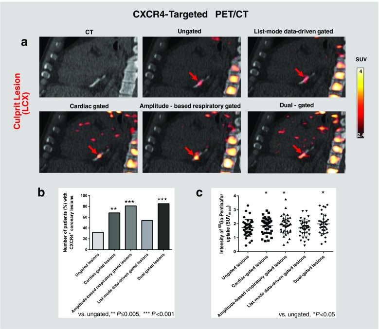
CXCR4 expression was analysed ex vivo in separately obtained arterial wall specimens. [Ga]Pentixafor PET/CT was performed in 37 patients after stent-based reperfusion for a first acute ST-segment elevation myocardial infarction. List-mode PET data were reconstructed to five different datasets using cardiac and/or respiratory gating. Guided by CT for localization, the PET signals of culprit and various groups of nonculprit coronary lesions were analysed and compared.
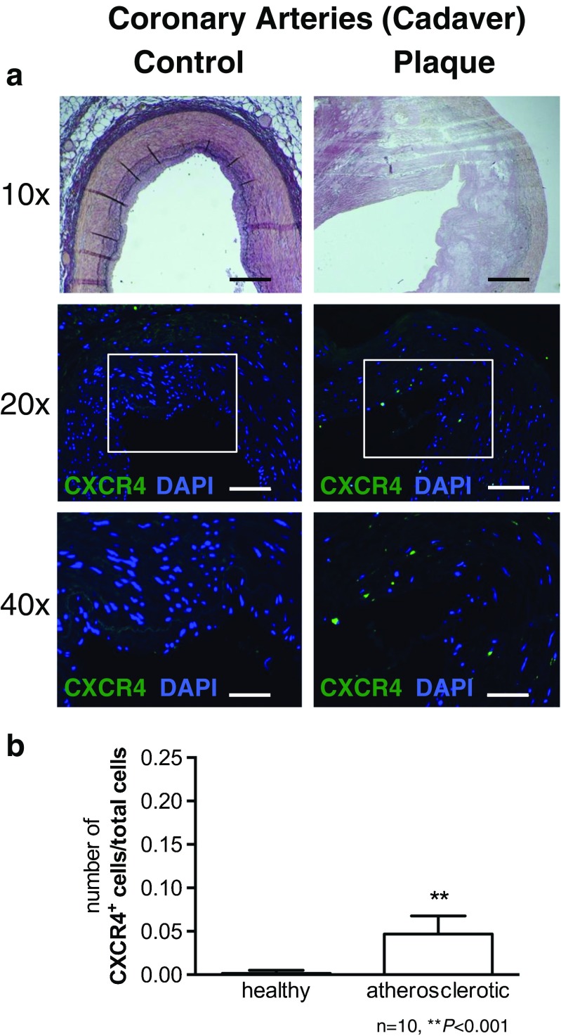
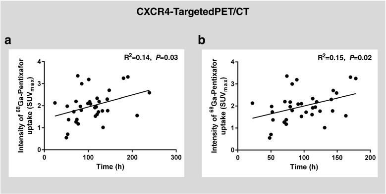
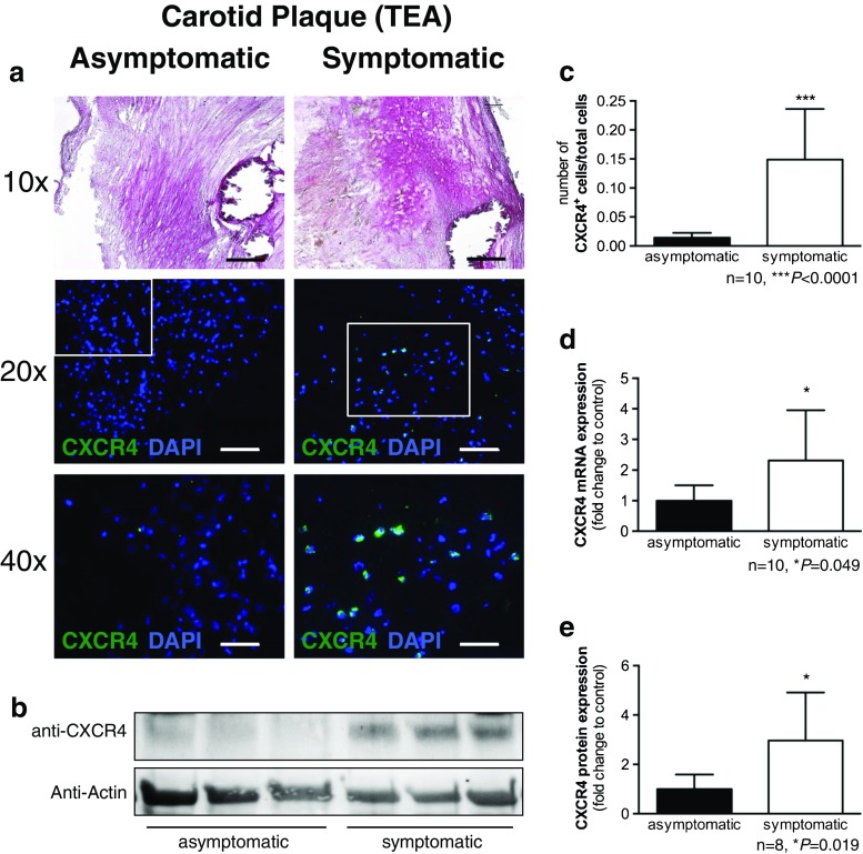
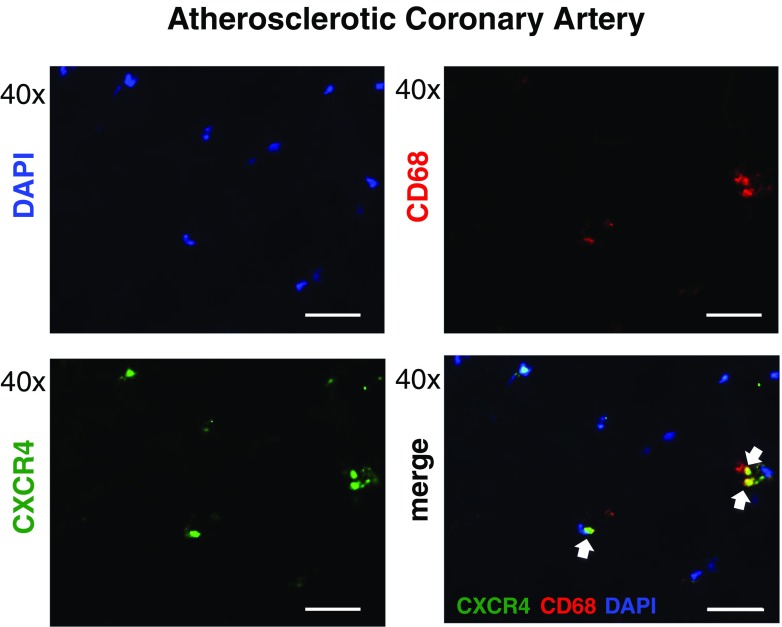
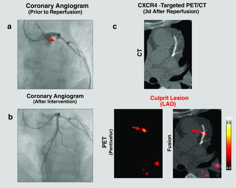
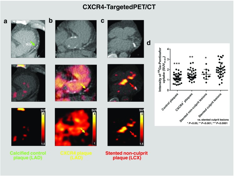
Ex vivo, CXCR4 was upregulated in atherosclerotic lesions, and mainly colocalized with CD68 inflammatory cells. In vivo, elevated CXCR4 expression was detected in culprit and nonculprit lesions, and the strongest CXCR4 PET signal (median SUV 1.96; interquartile range, IQR, 1.55-2.31) was observed in culprit coronary artery lesions. Stented nonculprit lesions (median SUV 1.45, IQR 1.23-1.88; P = 0.048) and hot spots in naive remote coronary segments (median SUV 1.34, IQR 1.23-1.74; P = 0.0005) showed significantly lower levels of CXCR4 expression. Dual cardiac/respiratory gating provided the strongest CXCR4 PET signal and the highest lesion detectability.
We demonstrated the basic feasibility of motion-corrected targeted PET/CT imaging of CXCR4 expression in coronary artery lesions, which was triggered by vessel wall inflammation but also by stent-induced injury. This novel methodology may serve as a platform for future diagnostic and therapeutic clinical studies targeting the biology of coronary atherosclerotic plaque.
趋化因子受体 CXCR4 是一种有前途的靶点,可用于对心血管疾病中 CXCR4 细胞类型(如炎症细胞)进行分子成像。我们推测,一种特定的 CXCR4 配体 [Ga]pentixafor 与新的运动校正技术相结合,将有助于通过运动校正靶向 PET/CT 对急性心肌梗死后小罪犯和非罪犯冠状动脉粥样硬化病变中的 CXCR4 表达进行体内特征分析。
在单独获得的动脉壁标本中分析 CXCR4 表达。对 37 例接受支架再灌注治疗的首次急性 ST 段抬高型心肌梗死患者进行 [Ga]Pentixafor PET/CT 检查。使用心脏和/或呼吸门控,对列表模式 PET 数据进行重建,得到五个不同的数据集。在 CT 引导下进行定位,分析和比较罪犯和各种非罪犯冠状动脉病变的 PET 信号。
在体内,CXCR4 在罪犯和非罪犯病变中均有升高,并且主要与 CD68 炎性细胞共定位。在体内,在罪犯和非罪犯病变中检测到 CXCR4 表达升高,在罪犯冠状动脉病变中观察到最强的 CXCR4 PET 信号(中位数 SUV1.96;四分位距,IQR,1.55-2.31)。支架内非罪犯病变(中位数 SUV1.45,IQR1.23-1.88;P=0.048)和未受累的远程冠状动脉节段的热点(中位数 SUV1.34,IQR1.23-1.74;P=0.0005)的 CXCR4 表达水平显著降低。双心脏/呼吸门控提供了最强的 CXCR4 PET 信号和最高的病变可检测性。
我们证明了运动校正靶向 PET/CT 成像在冠状动脉病变中 CXCR4 表达的基本可行性,这种方法是由血管壁炎症触发的,但也由支架诱导的损伤触发。这种新的方法学可能成为未来针对冠状动脉粥样硬化斑块生物学的诊断和治疗临床研究的平台。