Department of Radiology, University of Occupational and Environmental Health, Kitakyushu, Japan.
Department of Psychiatry, University of Occupational and Environmental Health, Kitakyushu, Japan.
Sci Rep. 2018 Jul 3;8(1):10054. doi: 10.1038/s41598-018-28300-5.
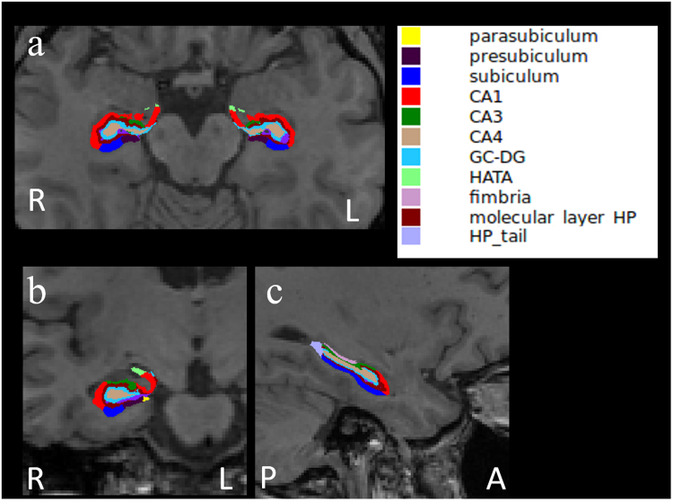
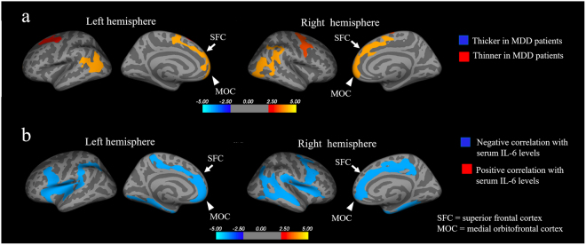
There is a growing body of evidence to support the involvement of proinflammatory cytokines in the pathophysiology of depression; however, no previous studies have examined the relationship between cytokines and the brain morphology of patients with major depressive disorder (MDD). We therefore evaluated the relationship between serum cytokine levels and cortical thinning during the first depressive episode in drug-naïve patients with MDD. We measured the serum cytokine levels (IL-1β, IL-6, IFN-γ, and TNFα), and whole-brain cortical thickness and hippocampal subfield volumes on brain magnetic resonance imaging (MRI) using surface-based morphometry in 40 patients with MDD and 47 healthy volunteers (controls). Only the serum IL-6 level was significantly higher in patients with MDD than in controls. The prefrontal cortex (PFC) thickness was significantly reduced in patients with MDD, and showed a significant inverse correlation with the serum IL-6 level. Although high serum IL-6 levels were correlated with reduced left subiculum and right CA1, CA3, CA4, GC-DG, subiculum, and whole hippocampus volumes, the presence or absence of MDD had no effect on the volume of any hippocampal subfields. Our results suggest that IL-6 may play a key role in the morphological changes in the PFC during the early stage of MDD.
越来越多的证据支持促炎细胞因子参与抑郁症的病理生理学;然而,以前的研究尚未检查细胞因子与重度抑郁症(MDD)患者的大脑形态之间的关系。因此,我们评估了在 MDD 药物初治患者的首次抑郁发作期间血清细胞因子水平与皮质变薄之间的关系。我们在 40 名 MDD 患者和 47 名健康志愿者(对照组)中使用基于表面的形态计量学测量了血清细胞因子水平(IL-1β、IL-6、IFN-γ 和 TNFα)以及全脑皮质厚度和海马亚区体积。与对照组相比,MDD 患者的血清 IL-6 水平显著升高。MDD 患者的前额叶皮质(PFC)厚度显著降低,并且与血清 IL-6 水平呈显著负相关。虽然高血清 IL-6 水平与左侧下托和右侧 CA1、CA3、CA4、GC-DG、下托和整个海马体体积减少相关,但 MDD 的存在与否对任何海马亚区的体积均无影响。我们的结果表明,IL-6 可能在 MDD 早期 PFC 的形态变化中起关键作用。