Department of Ophthalmology, Wakayama Medical University, 811-1 Kimiidera, Wakayama, 641-0012, Japan.
School of Ophthalmology and Optometry, Wenzhou Medical University, Zhejiang, China.
Cell Tissue Res. 2018 Nov;374(2):329-338. doi: 10.1007/s00441-018-2878-y. Epub 2018 Jul 4.
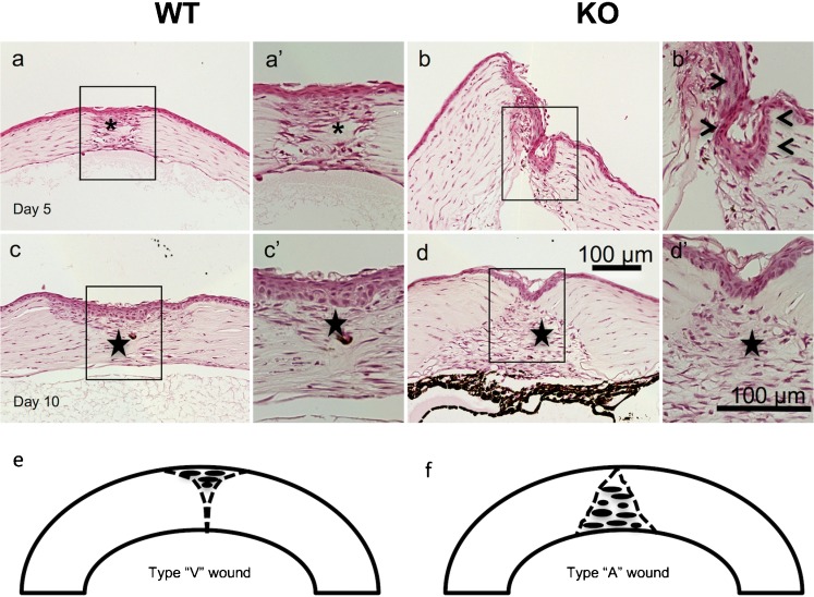
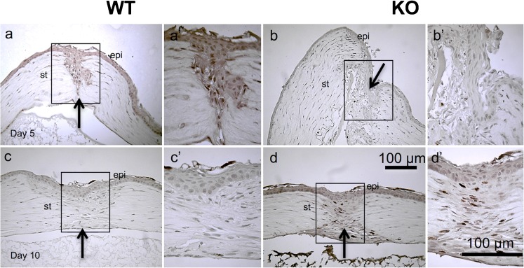
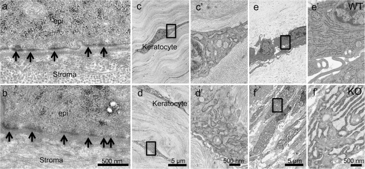
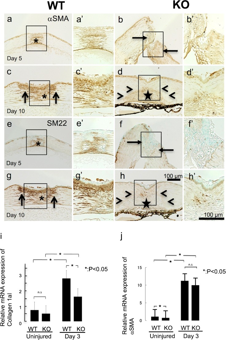
The present study attempts to elucidate the role of TRPV1 cation channel receptor on primary repair in an incision-wounded mouse cornea in vivo. Previous study revealed that blocking TRPV1 suppressed myofibroblast formation and expression of transforming growth factor β1 (TGFβ1) in cultured keratocytes or ocular fibroblasts. Male C57BL/6 (wild-type; WT) mice and male C57BL/6 Trpv1-null (KO) mice incurred a full-thickness incision injury (1.8 mm in length, limbus to limbus) in the central cornea of one eye with a surgical blade under general and topical anesthesia. The injury was not sutured. On days 0, 5, and 10, the eyes were enucleated, processed for histology, immunohistochemistry, and real-time RT-PCR gene expression analysis to evaluate the effects of the loss of TRPV1 on primary healing. Electron microscopy observation was also performed to know the effect of the loss of TRPV1 on ultrastructure of keratocytes. The results showed that the loss of Trpv1 gene delayed closure of corneal stromal incision with hindered myofibroblast transdifferentiation along with declines in the expression of collagen Ia1 and TGFβ1. Inflammatory cell infiltration was not affected by the loss of TRPV1. Ultrastructurally endoplasmic reticulum of TRPV1-null keratocytes was more extensively dilated as compared with WT keratocytes, suggesting an impairment of protein secretion by TRPV1-gene knockout. These results indicate that injury-related TRPV1 signal is involved in healing of stromal incision injury in a mouse cornea by selectively stimulating TGFβ-induced granulation tissue formation.
本研究试图阐明 TRPV1 阳离子通道受体在体内切口性小鼠角膜原发性修复中的作用。先前的研究表明,阻断 TRPV1 可抑制培养的角膜细胞或眼成纤维细胞中成肌纤维细胞的形成和转化生长因子 β1(TGFβ1)的表达。雄性 C57BL/6(野生型;WT)小鼠和雄性 C57BL/6 Trpv1 基因敲除(KO)小鼠在全身麻醉和局部麻醉下,用手术刀片在一只眼睛的中央角膜上造成全层切口损伤(长度 1.8 毫米,角膜缘到角膜缘)。损伤未缝合。在第 0、5 和 10 天,眼球被切除,进行组织学、免疫组织化学和实时 RT-PCR 基因表达分析,以评估 TRPV1 缺失对原发性愈合的影响。还进行了电子显微镜观察,以了解 TRPV1 缺失对角膜细胞超微结构的影响。结果表明,Trpv1 基因缺失延迟了角膜基质切口的闭合,伴有成肌纤维细胞转化的受阻,胶原 Ia1 和 TGFβ1 的表达下降。TRPV1 缺失对炎症细胞浸润没有影响。与 WT 角膜细胞相比,TRPV1 基因敲除的角膜细胞内质网扩张更为广泛,提示 TRPV1 基因敲除会损害蛋白质分泌。这些结果表明,与损伤相关的 TRPV1 信号通过选择性刺激 TGFβ 诱导的肉芽组织形成参与了小鼠角膜基质切口损伤的愈合。