Stem Cell Facility, (DBT-Centre of Excellence for Stem Cell Research), All India Institute of Medical Sciences, New Delhi, 110029, India.
Department of Pathology, All India Institute of Medical Sciences, New Delhi, India.
Stem Cell Res Ther. 2018 Jul 4;9(1):180. doi: 10.1186/s13287-018-0923-0.
Exosomes are nanovesicles (30-120 nm) of endosomal origin. These exosomes contain various functional proteins and RNAs that could be used for therapeutic purposes. Currently, having a standard method for exosome isolation retaining its biological properties with increased yield and purity is a major challenge. The most commonly used method is differential ultracentrifugation but it has its own disadvantages, which include high time consumption, low yield due to disruption of exosome integrity, and high protein contaminants. In this study, we have identified an improved method addressing these problems for exosome isolation using ultracentrifugation since it is cost-effective and used worldwide.
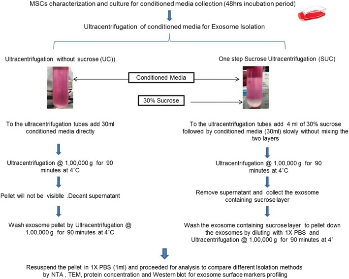
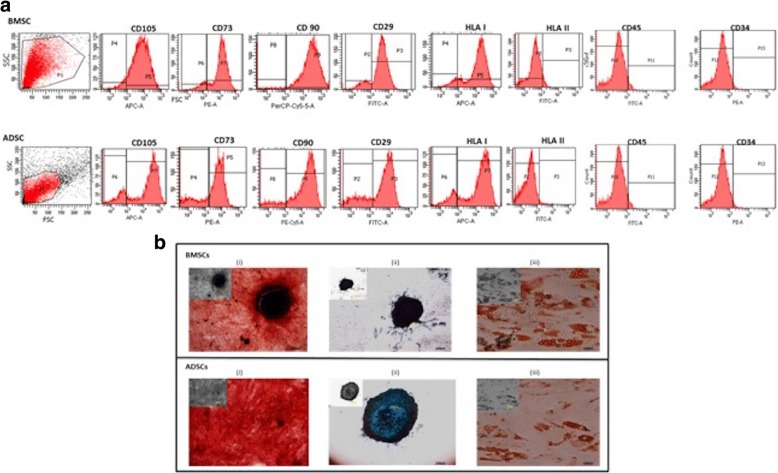
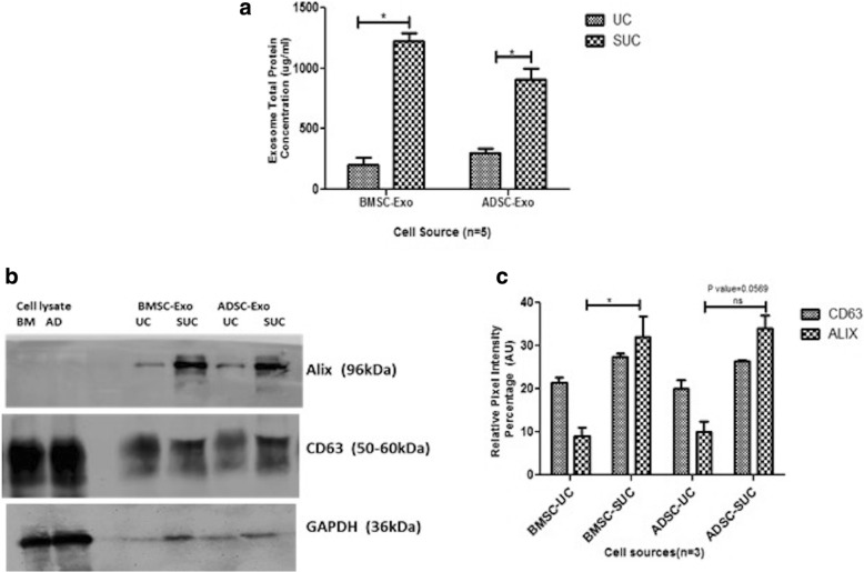
We have compared differential ultracentrifugation with the modified method called one-step sucrose cushion ultracentrifugation for exosome isolation. The conditioned serum-free media from human mesenchymal stem cells cultured for 48 h was collected for exosome isolation. The cellular debris was removed by centrifugation at 300g for 10 min, followed by centrifugation at 10,000g for 30 min to remove microvesicles. Equal volumes of pre-processed conditioned media were used for exosome isolation by direct ultracentrifugation and one-step sucrose cushion ultracentrifugation. The exosomes isolated using these methods were characterized for their size, morphology, concentration, and surface marker protein expression.
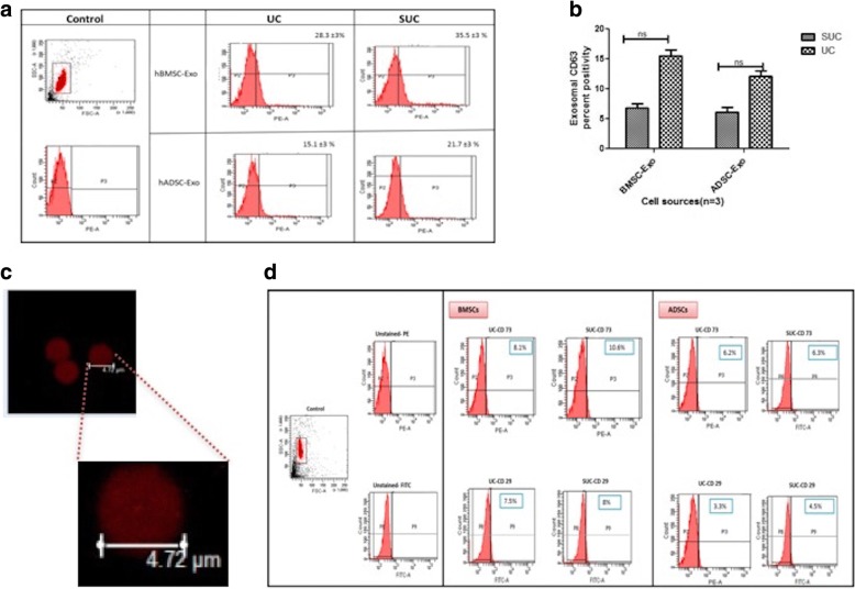
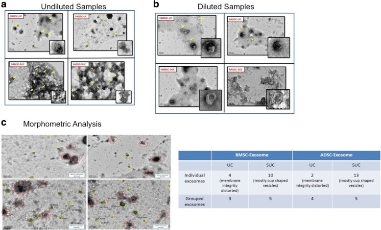
It was observed that the recovery of exosomes with cup-shaped morphology from one-step sucrose cushion ultracentrifugation was comparatively high as estimated by nanoparticle tracking analysis and electron microscopy. These results were confirmed by Western blotting and flow cytometry.
We conclude that this one-step sucrose cushion ultracentrifugation method provides an effective and reproducible potential standard method which could be used for various starting materials for isolating exosomes. We believe that this method will have a wide application in the field of extracellular vesicle research where exosome isolation with high yield and purity is an imperative step. Schematic representation of comparison of UC and SUC exosome isolation methods for tissue-specific human mesenchymal stem cells. The SUC isolation method yields a greater number of cup-shaped exosomes with a relatively homogenous population for mass-scale production of exosomes for downstream analysis.
SUC One-step sucrose cushion ultracentrifugation, UC Direct ultracentrifugation.
外泌体是起源于内体的纳米囊泡(30-120nm)。这些外泌体包含各种功能蛋白和 RNA,可用于治疗目的。目前,拥有一种标准的方法来分离保留其生物学特性的外泌体,同时提高产量和纯度,这是一个主要的挑战。最常用的方法是差速超速离心,但它有其自身的缺点,包括时间消耗长、由于外泌体完整性的破坏导致产量低以及蛋白质污染物含量高。在这项研究中,我们已经确定了一种改进的方法,用于使用超速离心法分离外泌体,因为它具有成本效益并且在全球范围内使用。
我们比较了差速超速离心法与改良的一步蔗糖垫超速离心法,用于分离外泌体。从培养 48 小时的人骨髓间充质干细胞的无血清条件培养基中收集外泌体进行分离。通过 300g 离心 10min 去除细胞碎片,然后通过 10,000g 离心 30min 去除微泡。使用预处理的条件培养基等体积,通过直接超速离心和一步蔗糖垫超速离心分离外泌体。使用这些方法分离的外泌体的大小、形态、浓度和表面标记蛋白表达进行了特征描述。
观察到一步蔗糖垫超速离心法从外泌体中回收杯状形态的外泌体的回收率相对较高,这是通过纳米颗粒跟踪分析和电子显微镜估计的。Western 印迹和流式细胞术证实了这一结果。
我们得出结论,这种一步蔗糖垫超速离心法提供了一种有效且可重复的潜在标准方法,可用于各种起始材料分离外泌体。我们相信,这种方法将在细胞外囊泡研究领域得到广泛应用,在外泌体产量和纯度高的情况下,这是一个必要的步骤。组织特异性人骨髓间充质干细胞 UC 和 SUC 外泌体分离方法比较示意图。SUC 分离方法产生了更多数量的杯状外泌体,具有相对同质的群体,可大规模生产外泌体进行下游分析。
SUC 一步蔗糖垫超速离心法,UC 直接超速离心法。