Departamento de Reproducción Animal, Instituto Nacional de Investigación y Tecnología Agraria y Alimentaria, Avenida Puerta de Hierro 12, local 10, 28040, Madrid, Spain.
Stem Cell Res Ther. 2018 Jul 4;9(1):178. doi: 10.1186/s13287-018-0933-y.
Recently, the capacity of mesenchymal stem/stromal cells (MSCs) to migrate into damaged tissues has been reported. For MSCs to be a promising tool for tissue engineering and cell and gene therapy, it is essential to know their migration ability according to their tissue of origin. However, little is known about the molecular mechanisms regulating porcine MSC chemotaxis. The aim of this study was to examine the migratory properties in an inflammatory environment of porcine MSC lines from different tissue origins: subcutaneous adipose tissue (SCA-MSCs), abdominal adipose tissue (AA-MSCs), dermal skin tissue (DS-MSCs) and peripheral blood (PB-MSCs).
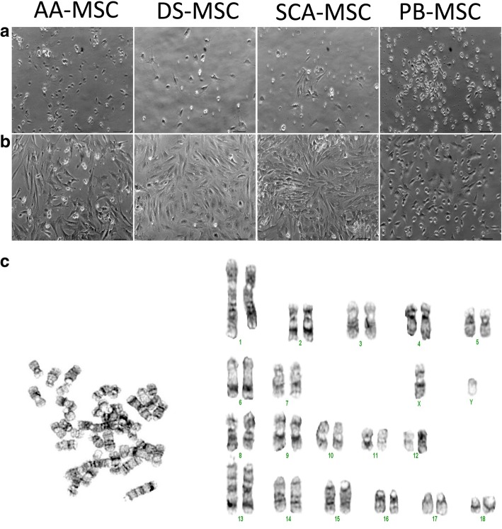
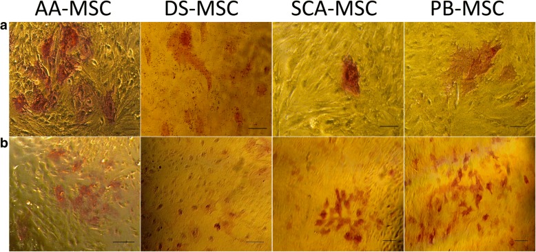
SCA-MSCs, AA-MSCs, DS-MSCs and PB-MSCs were isolated and analyzed in terms of morphological features, alkaline phosphatase activity, expression of cell surface and intracellular markers of pluripotency, proliferation, in vitro chondrogenic, osteogenic and adipogenic differentiation capacities, as well as their ability to migrate in response to inflammatory cytokines.
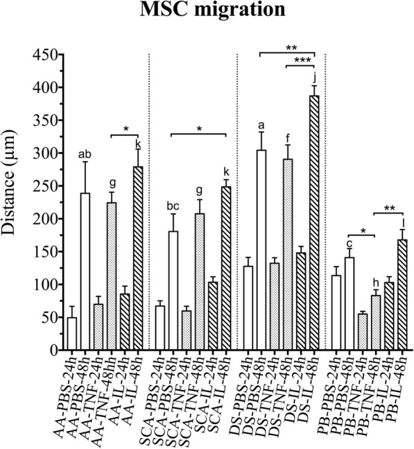
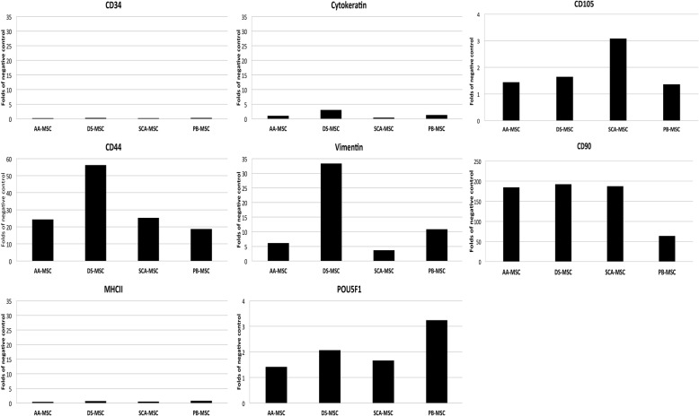
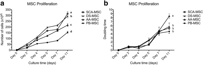
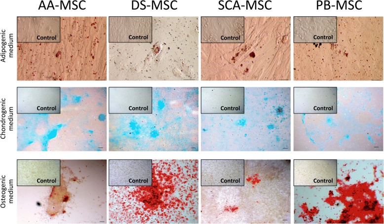
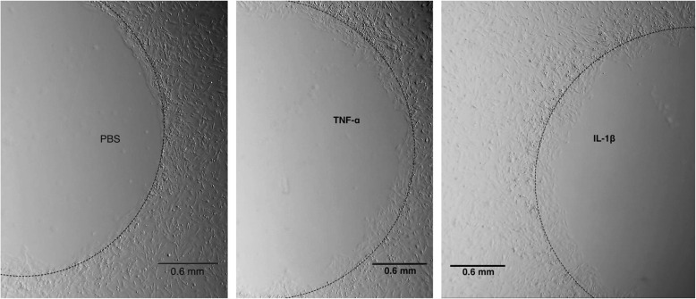
SCA-MSCs, AA-MSCs, DS-MSCs and PB-MSCs were isolated and showed plastic adhesion with a fibroblast-like morphology. All MSC lines were positive for CD44, CD105, CD90 and vimentin, characteristic markers of MSCs. The cytokeratin marker was also detected in DS-MSCs. No expression of MHCII or CD34 was detected in any of the four types of MSC. In terms of pluripotency features, all MSC lines expressed POU5F1 and showed alkaline phosphatase activity. SCA-MSCs had a higher growth rate compared to the rest of the cell lines, while the AA-MSC cell line had a longer population doubling time. All MSC lines cultured under adipogenic, chondrogenic and osteogenic conditions showed differentiation capacity to the previously mentioned mesodermal lineages. All MSC lines showed migration ability in an agarose drop assay. DS-MSCs migrated greater distances than the rest of the cell lines both in nonstimulated conditions and in the presence of the inflammatory cytokines TNF-α and IL-1β. SCA-MSCs and DS-MSCs increased their migration capacity in the presence of IL-1β as compared to PBS control.
This study describes the isolation and characterization of porcine cell lines from different tissue origin, with clear MSC properties. We show for the first time a comparative study of the migration capacity induced by inflammatory mediators of porcine MSCs of different tissue origin.
最近,间充质干细胞(MSCs)向损伤组织迁移的能力已被报道。为了使 MSCs 成为组织工程和细胞及基因治疗的有前途的工具,根据其组织来源了解其迁移能力至关重要。然而,对于调节猪 MSC 趋化性的分子机制知之甚少。本研究的目的是研究不同组织来源的猪 MSC 系在炎症环境中的迁移特性:皮下脂肪组织(SCA-MSCs)、腹部脂肪组织(AA-MSCs)、真皮组织(DS-MSCs)和外周血(PB-MSCs)。
分离 SCA-MSCs、AA-MSCs、DS-MSCs 和 PB-MSCs,并从形态特征、碱性磷酸酶活性、多能性细胞表面和细胞内标志物的表达、体外软骨形成、成骨和成脂分化能力以及对炎症细胞因子的迁移能力等方面进行分析。
分离出 SCA-MSCs、AA-MSCs、DS-MSCs 和 PB-MSCs,它们表现出贴壁生长的成纤维样形态。所有 MSC 系均表达 CD44、CD105、CD90 和波形蛋白,这些是 MSC 的特征标志物。DS-MSCs 也检测到细胞角蛋白标志物。四种 MSC 类型均未检测到 MHCII 或 CD34 的表达。在多能性特征方面,所有 MSC 系均表达 POU5F1 并表现出碱性磷酸酶活性。SCA-MSCs 的生长速度高于其余细胞系,而 AA-MSC 细胞系的倍增时间较长。在成脂、成软骨和成骨条件下培养的所有 MSC 系均表现出向先前提到的中胚层谱系分化的能力。所有 MSC 系在琼脂糖滴实验中均显示出迁移能力。在非刺激条件下以及在炎症细胞因子 TNF-α和 IL-1β存在的情况下,DS-MSCs 的迁移距离均大于其余细胞系。与 PBS 对照相比,SCA-MSCs 和 DS-MSCs 在存在 IL-1β的情况下增加了其迁移能力。
本研究描述了从不同组织来源分离和鉴定猪细胞系的方法,这些细胞系具有明确的 MSC 特性。我们首次比较研究了不同组织来源的猪 MSC 对炎症介质诱导的迁移能力。