• 文献检索
  • 文档翻译
  • 深度研究
  • 学术资讯
  • Suppr Zotero 插件Zotero 插件
  • 邀请有礼
  • 套餐&价格
  • 历史记录
应用&插件
Suppr Zotero 插件Zotero 插件浏览器插件Mac 客户端Windows 客户端微信小程序
定价
高级版会员购买积分包购买API积分包
服务
文献检索文档翻译深度研究API 文档MCP 服务
关于我们
关于 Suppr公司介绍联系我们用户协议隐私条款
关注我们

Suppr 超能文献

核心技术专利:CN118964589B侵权必究
粤ICP备2023148730 号-1Suppr @ 2026

文献检索

告别复杂PubMed语法,用中文像聊天一样搜索,搜遍4000万医学文献。AI智能推荐,让科研检索更轻松。

立即免费搜索

文件翻译

保留排版,准确专业,支持PDF/Word/PPT等文件格式,支持 12+语言互译。

免费翻译文档

深度研究

AI帮你快速写综述,25分钟生成高质量综述,智能提取关键信息,辅助科研写作。

立即免费体验

单细胞分析构建冠状动脉的早期祖细胞。

Single-cell analysis of early progenitor cells that build coronary arteries.

机构信息

Department of Biology, Stanford University, Stanford, CA, USA.

Program in Biophysics, Stanford University, Stanford, CA, USA.

出版信息

Nature. 2018 Jul;559(7714):356-362. doi: 10.1038/s41586-018-0288-7. Epub 2018 Jul 4.

DOI:10.1038/s41586-018-0288-7
PMID:29973725
原文链接:https://pmc.ncbi.nlm.nih.gov/articles/PMC6053322/
Abstract

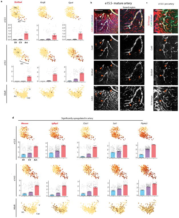
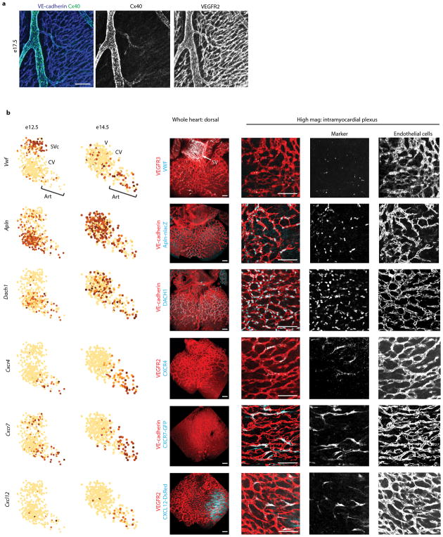
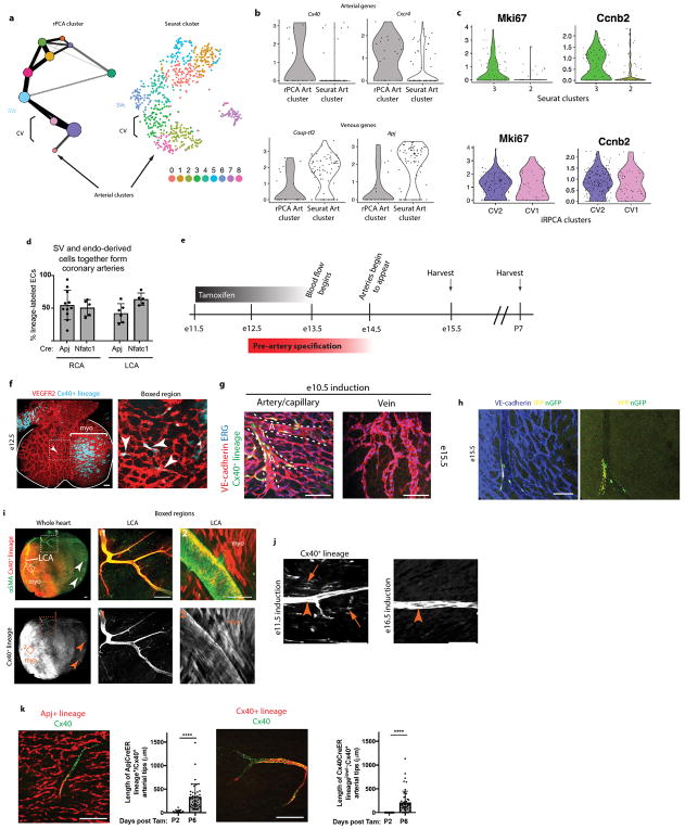
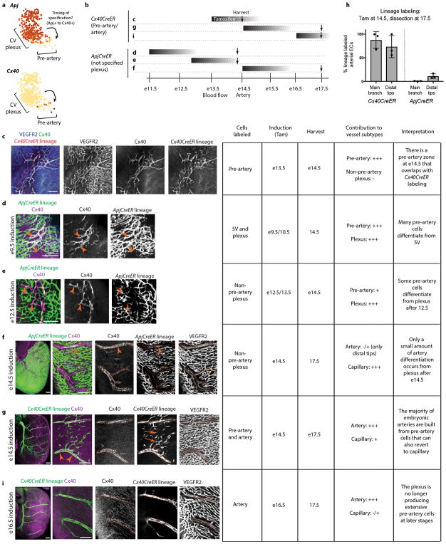
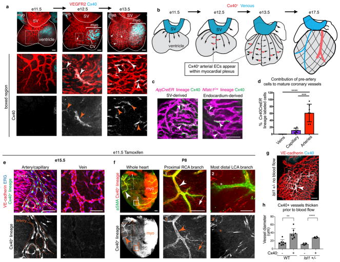
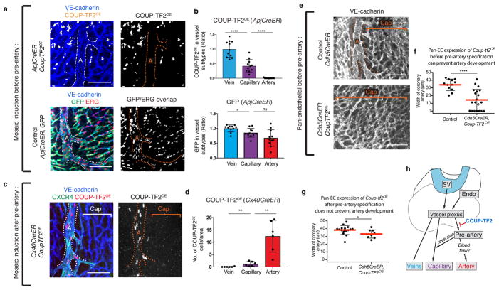
Arteries and veins are specified by antagonistic transcriptional programs. However, during development and regeneration, new arteries can arise from pre-existing veins through a poorly understood process of cell fate conversion. Here, using single-cell RNA sequencing and mouse genetics, we show that vein cells of the developing heart undergo an early cell fate switch to create a pre-artery population that subsequently builds coronary arteries. Vein cells underwent a gradual and simultaneous switch from venous to arterial fate before a subset of cells crossed a transcriptional threshold into the pre-artery state. Before the onset of coronary blood flow, pre-artery cells appeared in the immature vessel plexus, expressed mature artery markers, and decreased cell cycling. The vein-specifying transcription factor COUP-TF2 (also known as NR2F2) prevented plexus cells from overcoming the pre-artery threshold by inducing cell cycle genes. Thus, vein-derived coronary arteries are built by pre-artery cells that can differentiate independently of blood flow upon the release of inhibition mediated by COUP-TF2 and cell cycle factors.

摘要

动脉和静脉由拮抗的转录程序决定。然而,在发育和再生过程中,新的动脉可以通过一个尚未被充分了解的细胞命运转换过程从现有的静脉中产生。在这里,我们使用单细胞 RNA 测序和小鼠遗传学,表明发育中心脏的静脉细胞经历了早期的细胞命运转换,产生了一个前动脉群体,随后构建了冠状动脉。静脉细胞在亚群细胞跨越转录阈值进入前动脉状态之前,逐渐且同时从静脉命运向动脉命运转变。在冠状动脉血流开始之前,前动脉细胞出现在未成熟的血管丛中,表达成熟的动脉标记物,并减少细胞周期。静脉特异性转录因子 COUP-TF2(也称为 NR2F2)通过诱导细胞周期基因,阻止丛状细胞克服前动脉阈值。因此,静脉衍生的冠状动脉是由前动脉细胞构建的,这些细胞可以在 COUP-TF2 和细胞周期因子介导的抑制释放后独立于血流分化。

https://cdn.ncbi.nlm.nih.gov/pmc/blobs/bf3f/6053322/08da980578dc/nihms971464f5.jpg
https://cdn.ncbi.nlm.nih.gov/pmc/blobs/bf3f/6053322/0c40f503617c/nihms971464f6.jpg
https://cdn.ncbi.nlm.nih.gov/pmc/blobs/bf3f/6053322/4e116c4f9fa5/nihms971464f7.jpg
https://cdn.ncbi.nlm.nih.gov/pmc/blobs/bf3f/6053322/2411e86d0960/nihms971464f8.jpg
https://cdn.ncbi.nlm.nih.gov/pmc/blobs/bf3f/6053322/0816cc7770cc/nihms971464f9.jpg
https://cdn.ncbi.nlm.nih.gov/pmc/blobs/bf3f/6053322/8d088c0e99ad/nihms971464f10.jpg
https://cdn.ncbi.nlm.nih.gov/pmc/blobs/bf3f/6053322/036454bd8c63/nihms971464f11.jpg
https://cdn.ncbi.nlm.nih.gov/pmc/blobs/bf3f/6053322/8356bf34efb2/nihms971464f12.jpg
https://cdn.ncbi.nlm.nih.gov/pmc/blobs/bf3f/6053322/1f13acd0eb6f/nihms971464f13.jpg
https://cdn.ncbi.nlm.nih.gov/pmc/blobs/bf3f/6053322/34a23908bc9e/nihms971464f14.jpg
https://cdn.ncbi.nlm.nih.gov/pmc/blobs/bf3f/6053322/495084f81250/nihms971464f15.jpg
https://cdn.ncbi.nlm.nih.gov/pmc/blobs/bf3f/6053322/8bf2a3300f5c/nihms971464f1.jpg
https://cdn.ncbi.nlm.nih.gov/pmc/blobs/bf3f/6053322/4ed33026684d/nihms971464f2.jpg
https://cdn.ncbi.nlm.nih.gov/pmc/blobs/bf3f/6053322/2ccbcd1d5edf/nihms971464f3.jpg
https://cdn.ncbi.nlm.nih.gov/pmc/blobs/bf3f/6053322/b6f1119848dd/nihms971464f4.jpg
https://cdn.ncbi.nlm.nih.gov/pmc/blobs/bf3f/6053322/08da980578dc/nihms971464f5.jpg

相似文献

1
Single-cell analysis of early progenitor cells that build coronary arteries.单细胞分析构建冠状动脉的早期祖细胞。
Nature. 2018 Jul;559(7714):356-362. doi: 10.1038/s41586-018-0288-7. Epub 2018 Jul 4.
2
Genomic Characterization of Endothelial Enhancers Reveals a Multifunctional Role for NR2F2 in Regulation of Arteriovenous Gene Expression.内皮增强子的基因组特征揭示了 NR2F2 在调节动静脉基因表达中的多功能作用。
Circ Res. 2020 Mar 27;126(7):875-888. doi: 10.1161/CIRCRESAHA.119.316075. Epub 2020 Feb 17.
3
Suppression of Notch signalling by the COUP-TFII transcription factor regulates vein identity.COUP-TFII转录因子对Notch信号通路的抑制作用调控静脉特性。
Nature. 2005 May 5;435(7038):98-104. doi: 10.1038/nature03511.
4
Venous Endothelial Marker COUP-TFII Regulates the Distinct Pathologic Potentials of Adult Arteries and Veins.静脉内皮标志物COUP-TFII调节成年动脉和静脉的不同病理潜能。
Sci Rep. 2015 Nov 5;5:16193. doi: 10.1038/srep16193.
5
SoxF factors and Notch regulate nr2f2 gene expression during venous differentiation in zebrafish.SoxF 因子和 Notch 在斑马鱼静脉分化过程中调节 nr2f2 基因的表达。
Dev Biol. 2014 Jun 15;390(2):116-25. doi: 10.1016/j.ydbio.2014.03.018. Epub 2014 Mar 31.
6
Arterial and venous progenitors of the major axial vessels originate at distinct locations.主要轴向血管的动脉和静脉祖细胞起源于不同的位置。
Dev Cell. 2013 Apr 29;25(2):196-206. doi: 10.1016/j.devcel.2013.03.017.
7
Lymphatic vessels arise from specialized angioblasts within a venous niche.淋巴管由静脉龛内的专门血管母细胞产生。
Nature. 2015 Jun 4;522(7554):56-61. doi: 10.1038/nature14425.
8
Fluid shear stress induces arterial differentiation of endothelial progenitor cells.流体剪切应力诱导内皮祖细胞向动脉方向分化。
J Appl Physiol (1985). 2009 Jan;106(1):203-11. doi: 10.1152/japplphysiol.00197.2008. Epub 2008 Nov 6.
9
Arterial-venous endothelial cell fate is related to vascular endothelial growth factor and Notch status during human bone mesenchymal stem cell differentiation.在人骨髓间充质干细胞分化过程中,动静脉内皮细胞命运与血管内皮生长因子和Notch状态相关。
FEBS Lett. 2008 Aug 20;582(19):2957-64. doi: 10.1016/j.febslet.2008.07.031. Epub 2008 Jul 29.
10
Limited Regeneration Potential with Minimal Epicardial Progenitor Conversions in the Neonatal Mouse Heart after Injury.损伤后新生鼠心脏心外膜祖细胞转化为心肌细胞的能力有限,再生潜能有限。
Cell Rep. 2019 Jul 2;28(1):190-201.e3. doi: 10.1016/j.celrep.2019.06.003.

引用本文的文献

1
Combined Pharmacological Conditioning of Endothelial Cells for Improved Vascular Graft Endothelialization.联合药物调节内皮细胞以改善血管移植物内皮化
Int J Mol Sci. 2025 Jul 25;26(15):7183. doi: 10.3390/ijms26157183.
2
COUP-TFII-mediated reprogramming of the vascular endothelium counteracts tumor immune evasion.COUP-TFII介导的血管内皮重编程可对抗肿瘤免疫逃逸。
Nat Commun. 2025 Aug 12;16(1):7457. doi: 10.1038/s41467-025-62399-1.
3
Feto-placental blood vessel development.胎儿-胎盘血管发育

本文引用的文献

1
Shear-induced Notch-Cx37-p27 axis arrests endothelial cell cycle to enable arterial specification.剪切诱导的 Notch-Cx37-p27 轴使内皮细胞周期停滞,从而实现动脉特化。
Nat Commun. 2017 Dec 15;8(1):2149. doi: 10.1038/s41467-017-01742-7.
2
NOTCH1 is a mechanosensor in adult arteries.NOTCH1 是成体动脉中的机械感受器。
Nat Commun. 2017 Nov 20;8(1):1620. doi: 10.1038/s41467-017-01741-8.
3
Human haematopoietic stem cell lineage commitment is a continuous process.人类造血干细胞谱系定向分化是一个连续的过程。
Development. 2025 Jun 1;152(11). doi: 10.1242/dev.204838. Epub 2025 Jun 2.
4
Metabolic and Immune Crosstalk in Cardiovascular Disease.心血管疾病中的代谢与免疫相互作用
Circ Res. 2025 May 23;136(11):1433-1453. doi: 10.1161/CIRCRESAHA.125.325496. Epub 2025 May 22.
5
Dissecting endothelial cell heterogeneity with new tools.用新工具剖析内皮细胞异质性。
Cell Regen. 2025 Mar 23;14(1):10. doi: 10.1186/s13619-025-00223-3.
6
CXCL12 drives natural variation in coronary artery anatomy across diverse populations.CXCL12驱动不同人群冠状动脉解剖结构的自然变异。
Cell. 2025 Apr 3;188(7):1784-1806.e22. doi: 10.1016/j.cell.2025.02.005. Epub 2025 Mar 5.
7
Monocyte-Derived cxcl12 Guides a Directional Migration of Blood Vessels in Zebrafish.单核细胞衍生的趋化因子CXCL12引导斑马鱼血管的定向迁移。
Arterioscler Thromb Vasc Biol. 2025 Mar;45(3):386-397. doi: 10.1161/ATVBAHA.124.321588. Epub 2025 Jan 23.
8
Evaluating the transcriptional regulators of arterial gene expression via a catalogue of characterized arterial enhancers.通过已鉴定的动脉增强子目录评估动脉基因表达的转录调节因子。
Elife. 2025 Jan 17;14:e102440. doi: 10.7554/eLife.102440.
9
Dynamic behavior and lineage plasticity of the pulmonary venous endothelium.肺静脉内皮细胞的动态行为和谱系可塑性。
Nat Cardiovasc Res. 2024 Dec;3(12):1584-1600. doi: 10.1038/s44161-024-00573-2. Epub 2024 Dec 9.
10
Differential vegfc expression dictates lymphatic response during zebrafish heart development and regeneration.差异表达的 vegfc 决定斑马鱼心脏发育和再生过程中的淋巴管反应。
Development. 2024 Nov 15;151(22). doi: 10.1242/dev.202947. Epub 2024 Nov 19.
Nat Cell Biol. 2017 Apr;19(4):271-281. doi: 10.1038/ncb3493. Epub 2017 Mar 20.
4
An atlas of transcriptional, chromatin accessibility, and surface marker changes in human mesoderm development.人类中胚层发育中转录组、染色质可及性和表面标记变化的图谱。
Sci Data. 2016 Dec 20;3:160109. doi: 10.1038/sdata.2016.109.
5
Coronary Artery Development: Progenitor Cells and Differentiation Pathways.冠状动脉发育:祖细胞与分化途径
Annu Rev Physiol. 2017 Feb 10;79:1-19. doi: 10.1146/annurev-physiol-022516-033953. Epub 2016 Dec 9.
6
MEF2 transcription factors are key regulators of sprouting angiogenesis.MEF2转录因子是出芽血管生成的关键调节因子。
Genes Dev. 2016 Oct 15;30(20):2297-2309. doi: 10.1101/gad.290619.116.
7
Endothelial cells are progenitors of cardiac pericytes and vascular smooth muscle cells.内皮细胞是心脏周细胞和血管平滑肌细胞的前体细胞。
Nat Commun. 2016 Aug 12;7:12422. doi: 10.1038/ncomms12422.
8
Cellular Taxonomy of the Mouse Striatum as Revealed by Single-Cell RNA-Seq.单细胞RNA测序揭示的小鼠纹状体细胞分类学
Cell Rep. 2016 Jul 26;16(4):1126-1137. doi: 10.1016/j.celrep.2016.06.059. Epub 2016 Jul 14.
9
Mapping the Pairwise Choices Leading from Pluripotency to Human Bone, Heart, and Other Mesoderm Cell Types.描绘从多能性到人类骨骼、心脏及其他中胚层细胞类型的成对选择图谱。
Cell. 2016 Jul 14;166(2):451-467. doi: 10.1016/j.cell.2016.06.011.
10
Endocardium Minimally Contributes to Coronary Endothelium in the Embryonic Ventricular Free Walls.心内膜对胚胎心室游离壁的冠状动脉内皮的贡献极小。
Circ Res. 2016 Jun 10;118(12):1880-93. doi: 10.1161/CIRCRESAHA.116.308749. Epub 2016 Apr 7.