Xu Hui, Wang Xiaocui, Chen Zhen, Bai Guanghui, Yin Bo, Wang Shan, Sun Chuanzhu, Gan Shuoqiu, Wang Zhuonan, Cao Jieli, Niu Xuan, Shao Meihua, Gu Chenghui, Hu Liuxun, Ye Limei, Li Dandong, Yan Zhihan, Zhang Ming, Bai Lijun
The Key Laboratory of Biomedical Information Engineering, Ministry of Education, Department of Biomedical Engineering, School of Life Science and Technology, Xi'an Jiaotong University, Xi'an, China.
Department of Medical Imaging, The First Affiliated Hospital of Xi'an Jiaotong University, Xi'an, China.
Front Neurol. 2018 Jun 19;9:467. doi: 10.3389/fneur.2018.00467. eCollection 2018.
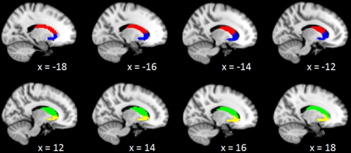
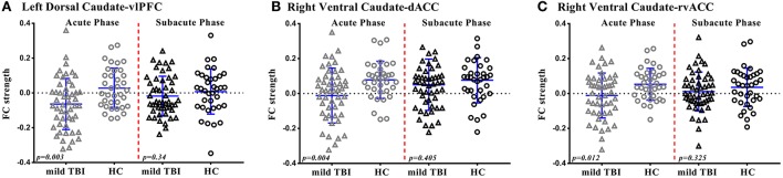
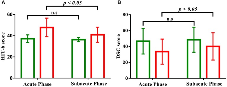
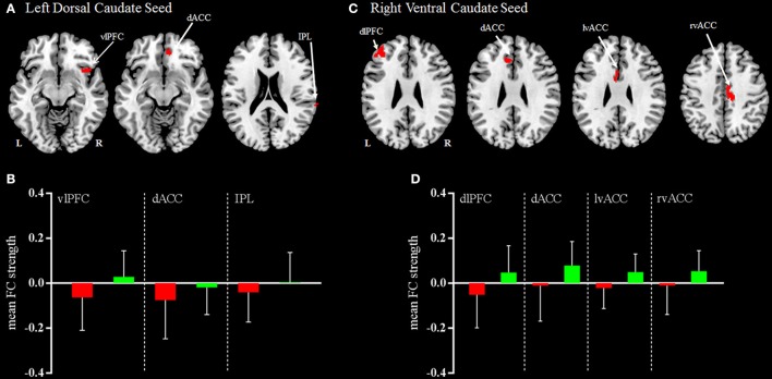
Mild traumatic brain injury (mild TBI) is associated with dysfunctional brain network and accumulating evidence is pointing to the caudate as a vulnerable hub region. However, little is known about the longitudinal changes in the caudate-based resting-state functional connectivity following mild TBI. In the current study, 50 patients with mild TBI received resting-state functional magnetic resonance imaging as well as neuropsychological assessments within 7 days post-injury (acute phase) and 1 month later (subacute phase). Thirty-six age- and gender- matched healthy controls underwent the same protocol. The caudate was segmented into the dorsal and ventral sub-regions based on their related functionally distinct neural circuits and separate functional connectivity was investigated. Results indicated that patients with mild TBI at acute phase exhibited reduced left dorsal caudate-based functional connectivity with ventral lateral prefrontal cortex, dorsal anterior cingulate cortex, and inferior parietal lobule, which mainly distributed in the cognitive control network, and reduced right ventral caudate-based functional connectivity with the dorsal lateral prefrontal cortex, dorsal anterior cingulate cortex (dACC), and bilateral ventral anterior cingulate cortex (vACC), which mainly distributed in the executive network and emotional processing network. Furthermore, patients with mild TBI presented the reduced functional connectivity between the left dorsal caudate and the ventral lateral prefrontal cortex (vlPFC) compared with healthy controls at acute phase while this difference became no significance and return to the normal level following 1 month post-injury subacute phase. Similarly, the functional connectivity between the right ventral caudate and anterior cingulate cortex (both dorsal and ventral part) showed the reduced strength in patients compared with healthy controls only at the acute phase but presented no significant difference at subacute phase following mild TBI. Along the same line, patients with mild TBI presented the impaired performance on the information processing speed and more complaints on the pain impact index at acute phase compared with healthy controls but showed no significant difference at the follow-up 1 month post-injury subacute phase. The longitudinal changes of caudate-based dysfunction connectivity could serve as a neuroimaging biomarker following patients with mild TBI, with the evidence that the abnormal caudate-based functional connectivity at acute phase have returned to the normal level accompanying with the recovery of the neuropsychological syndromes following patients with mild TBI at subacute phase.
轻度创伤性脑损伤(轻度TBI)与脑网络功能失调有关,越来越多的证据表明尾状核是一个易损枢纽区域。然而,关于轻度TBI后基于尾状核的静息态功能连接的纵向变化知之甚少。在本研究中,50例轻度TBI患者在受伤后7天内(急性期)和1个月后(亚急性期)接受了静息态功能磁共振成像以及神经心理学评估。36名年龄和性别匹配的健康对照者接受了相同的检查方案。根据尾状核相关的功能不同的神经回路,将尾状核分为背侧和腹侧子区域,并分别研究其功能连接。结果表明,急性期轻度TBI患者左侧基于背侧尾状核与腹外侧前额叶皮层、背侧前扣带回皮层和顶下小叶的功能连接减少,这些区域主要分布在认知控制网络中;右侧基于腹侧尾状核与背外侧前额叶皮层、背侧前扣带回皮层(dACC)和双侧腹侧前扣带回皮层(vACC)的功能连接减少,这些区域主要分布在执行网络和情绪处理网络中。此外,急性期轻度TBI患者左侧背侧尾状核与腹外侧前额叶皮层(vlPFC)之间的功能连接与健康对照相比减少,而这种差异在受伤后1个月的亚急性期变得不显著并恢复到正常水平。同样,右侧腹侧尾状核与前扣带回皮层(背侧和腹侧部分)之间的功能连接强度仅在急性期患者与健康对照相比降低,但在轻度TBI后的亚急性期无显著差异。同样,急性期轻度TBI患者与健康对照相比,在信息处理速度方面表现受损,在疼痛影响指数方面有更多主诉,但在受伤后1个月的亚急性期随访时无显著差异。基于尾状核的功能连接功能障碍的纵向变化可作为轻度TBI患者的神经影像学生物标志物,有证据表明急性期基于尾状核的异常功能连接在轻度TBI患者亚急性期随着神经心理学综合征的恢复已恢复到正常水平。