Huang Haiqing, Nguyen Peter T, Schwab Nadine A, Tanner Jared J, Price Catherine C, Ding Mingzhou
J. Crayton Pruitt Family Department of Biomedical Engineering, University of FloridaGainesville, FL, USA.
Department of Clinical and Health Psychology, University of FloridaGainesville, FL, USA.
Front Aging Neurosci. 2017 Apr 4;9:91. doi: 10.3389/fnagi.2017.00091. eCollection 2017.
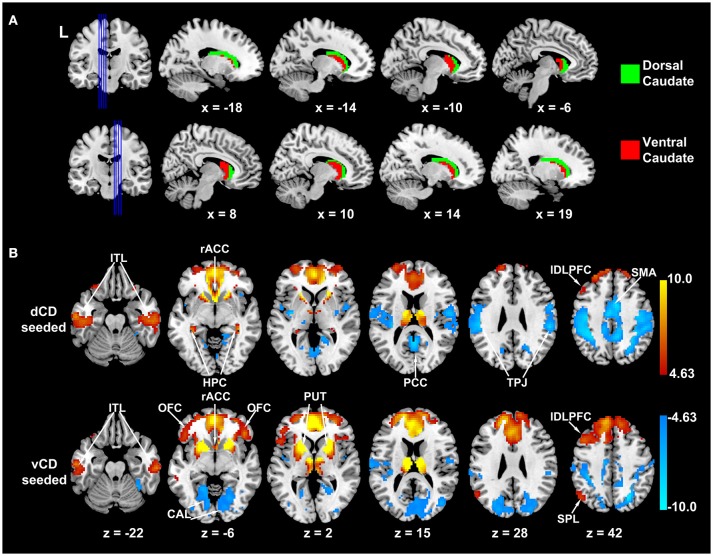
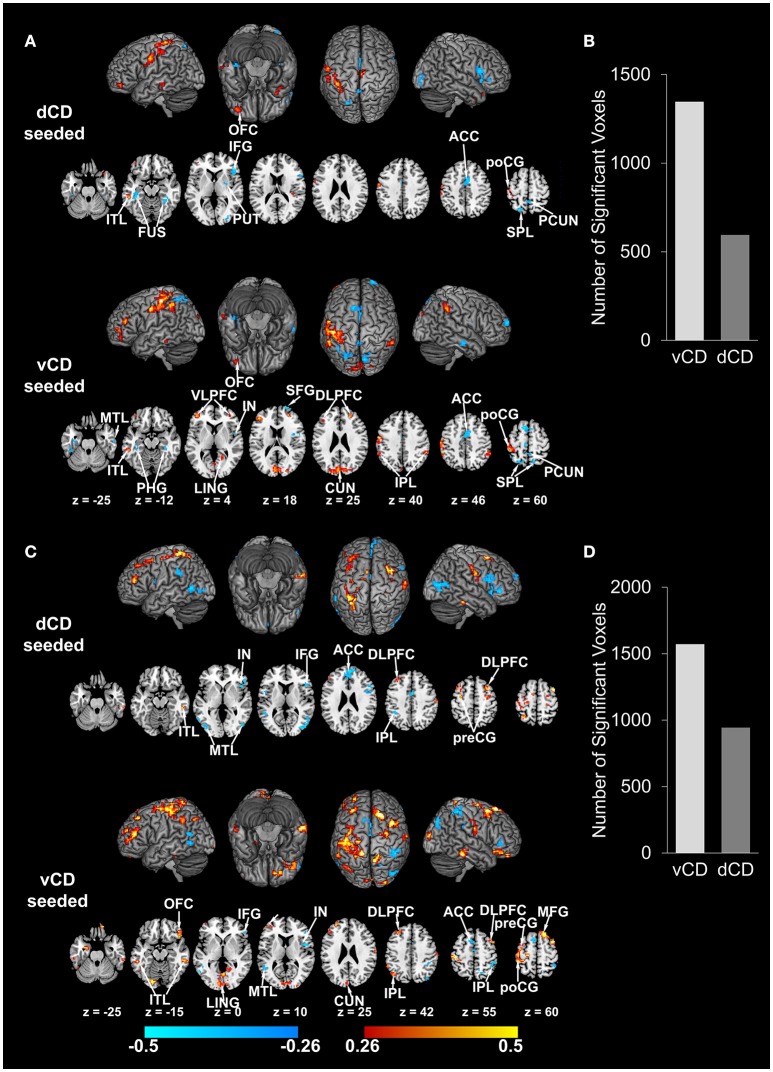
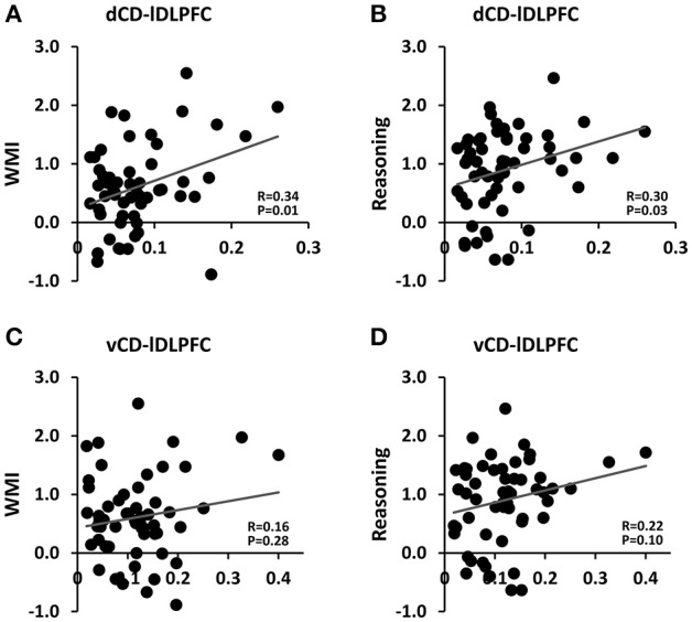
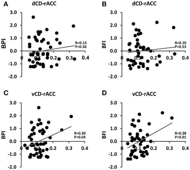
The caudate nucleus plays important roles in cognition and affect. Depending on associated connectivity and function, the caudate can be further divided into dorsal and ventral aspects. Dorsal caudate, highly connected to dorsolateral prefrontal cortex (DLPFC), is implicated in executive function and working memory; ventral caudate, more interconnected with the limbic system, is implicated in affective functions such as pain processing. Clinically, certain brain disorders are known to differentially impact dorsal and ventral caudate. Thus, precise parcellation of caudate has both basic and clinical neuroscience significance. In young adults, past work has combined resting-state fMRI functional connectivity with clustering algorithms to define dorsal and ventral caudate. Whether the same approach is effective in older adults and how to validate the parcellation results have not been considered. We addressed these problems by obtaining resting-state fMRI data from 56 older non-demented adults (age: 69.07 ± 5.92 years and MOCA: 25.71 ± 2.46) along with a battery of cognitive and clinical assessments. Connectivity from each voxel of caudate to the rest of the brain was computed using cross correlation. Applying the K-means clustering algorithm to the connectivity patterns with = 2 yielded two substructures within caudate, which agree well with previously reported dorsal and ventral divisions of caudate. Furthermore, dorsal-caudate-seeded functional connectivity was shown to be more strongly associated with working memory and fluid reasoning composite scores, whereas ventral-caudate-seeded functional connectivity more strongly associated with pain and fatigue severity. These results demonstrate that dorsal and ventral caudate can be reliably identified by combining resting-state fMRI and clustering algorithms in older adults.
尾状核在认知和情感方面发挥着重要作用。根据相关的连接性和功能,尾状核可进一步分为背侧和腹侧部分。背侧尾状核与背外侧前额叶皮层(DLPFC)高度连接,与执行功能和工作记忆有关;腹侧尾状核与边缘系统的相互连接更多,与诸如疼痛处理等情感功能有关。临床上,已知某些脑部疾病会对背侧和腹侧尾状核产生不同影响。因此,尾状核的精确分割具有基础和临床神经科学意义。在年轻人中,过去的研究将静息态功能磁共振成像(fMRI)功能连接性与聚类算法相结合来定义背侧和腹侧尾状核。但尚未考虑同样的方法在老年人中是否有效以及如何验证分割结果。我们通过获取56名非痴呆老年人(年龄:69.07±5.92岁,蒙特利尔认知评估量表(MOCA)得分:25.71±2.46)的静息态fMRI数据以及一系列认知和临床评估来解决这些问题。使用互相关计算尾状核每个体素与大脑其他部分的连接性。将K均值聚类算法应用于连接模式(k = 2),在尾状核内产生了两个子结构,这与先前报道的尾状核背侧和腹侧划分非常吻合。此外,以背侧尾状核为种子点的功能连接性与工作记忆和流体推理综合得分的相关性更强,而以腹侧尾状核为种子点的功能连接性与疼痛和疲劳严重程度的相关性更强。这些结果表明,在老年人中,通过结合静息态fMRI和聚类算法可以可靠地识别背侧和腹侧尾状核。