Balas Mihaela, Dumitrache Florian, Badea Madalina Andreea, Fleaca Claudiu, Badoi Anca, Tanasa Eugenia, Dinischiotu Anca
Department of Biochemistry and Molecular Biology, University of Bucharest, 91⁻95 Splaiul Independenţei, 050095 Bucharest, sector 5, Romania.
National Institute for Lasers, Plasma and Radiation Physics (NILPRP), Atomistilor 409, 077125 Magurele, Romania.
Nanomaterials (Basel). 2018 Jul 5;8(7):495. doi: 10.3390/nano8070495.
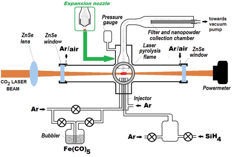
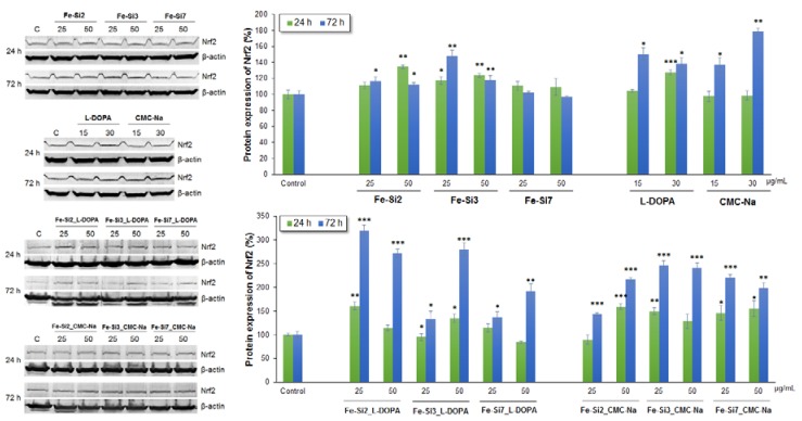
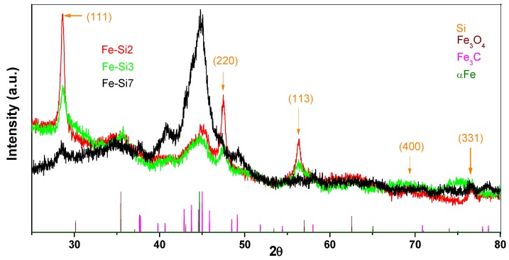
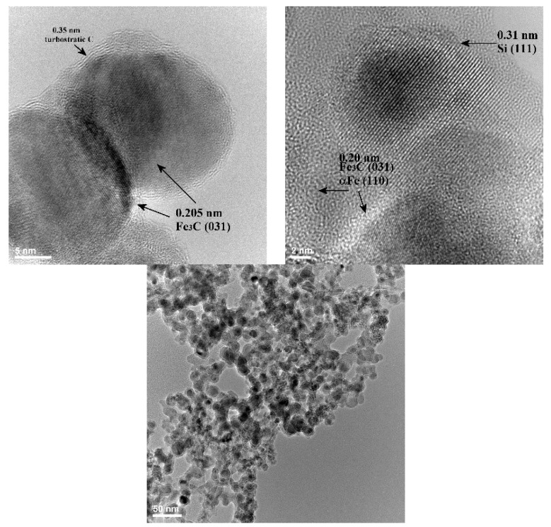
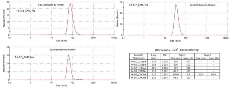
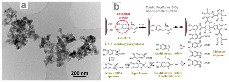
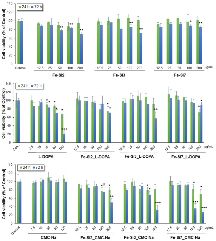
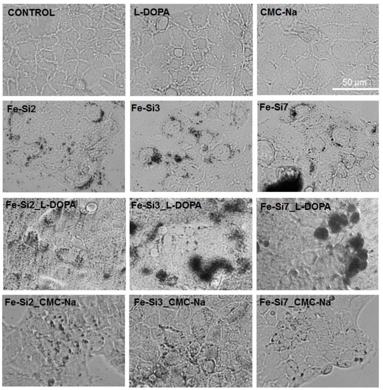
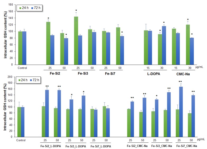
Magnetic nanoparticles offer multiple utilization possibilities in biomedicine. In this context, the interaction with cellular structures and their biological effects need to be understood and controlled for clinical safety. New magnetic nanoparticles containing metallic/carbidic iron and elemental silicon phases were synthesized by laser pyrolysis using Fe(CO)₅ vapors and SiH₄ gas as Fe and Si precursors, then passivated and coated with biocompatible agents, such as l-3,4-dihydroxyphenylalanine (l-DOPA) and sodium carboxymethyl cellulose (CMC-Na). The resulting magnetic nanoparticles were characterized by XRD, EDS, and TEM techniques. To evaluate their biocompatibility, doses ranging from 0⁻200 µg/mL hybrid Fe-Si nanoparticles were exposed to Caco2 cells for 24 and 72 h. Doses below 50 μg/mL of both l-DOPA and CMC-Na-coated Fe-Si nanoparticles induced no significant changes of cellular viability or membrane integrity. The cellular internalization of nanoparticles was dependent on their dispersion in culture medium and caused some changes of F-actin filaments organization after 72 h. However, reactive oxygen species were generated after exposure to 25 and 50 μg/mL of both Fe-Si nanoparticles types, inducing the increase of intracellular glutathione level and activation of transcription factor Nrf2. At nanoparticles doses below 50 μg/mL, Caco2 cells were able to counteract the oxidative stress by activating the cellular protection mechanisms. We concluded that in vitro biological responses to coated hybrid Fe-Si nanoparticles depended on particle synthesis conditions, surface coating, doses and incubation time.
磁性纳米颗粒在生物医学领域具有多种应用可能性。在此背景下,为确保临床安全性,需要了解并控制其与细胞结构的相互作用及其生物学效应。通过激光热解,以五羰基铁蒸汽(Fe(CO)₅)和硅烷气体(SiH₄)作为铁和硅的前驱体,合成了含有金属/碳化物铁相和元素硅相的新型磁性纳米颗粒,然后用生物相容性试剂(如L-3,4-二羟基苯丙氨酸(L-DOPA)和羧甲基纤维素钠(CMC-Na))进行钝化和包覆。通过X射线衍射(XRD)、能谱分析(EDS)和透射电子显微镜(TEM)技术对所得磁性纳米颗粒进行了表征。为评估其生物相容性,将浓度范围为0⁻200μg/mL的混合铁硅纳米颗粒暴露于Caco2细胞24小时和72小时。L-DOPA和CMC-Na包覆的铁硅纳米颗粒浓度低于50μg/mL时,细胞活力或膜完整性均未发生显著变化。纳米颗粒的细胞内化取决于其在培养基中的分散情况,72小时后会导致F-肌动蛋白丝组织发生一些变化。然而,两种类型的铁硅纳米颗粒在暴露于25μg/mL和50μg/mL时均会产生活性氧,导致细胞内谷胱甘肽水平升高并激活转录因子Nrf2。在纳米颗粒浓度低于50μg/mL时,Caco2细胞能够通过激活细胞保护机制来抵抗氧化应激。我们得出结论,体外对包覆的混合铁硅纳米颗粒的生物学反应取决于颗粒的合成条件、表面包覆、剂量和孵育时间。