• 文献检索
  • 文档翻译
  • 深度研究
  • 学术资讯
  • Suppr Zotero 插件Zotero 插件
  • 邀请有礼
  • 套餐&价格
  • 历史记录
应用&插件
Suppr Zotero 插件Zotero 插件浏览器插件Mac 客户端Windows 客户端微信小程序
定价
高级版会员购买积分包购买API积分包
服务
文献检索文档翻译深度研究API 文档MCP 服务
关于我们
关于 Suppr公司介绍联系我们用户协议隐私条款
关注我们

Suppr 超能文献

核心技术专利:CN118964589B侵权必究
粤ICP备2023148730 号-1Suppr @ 2026

文献检索

告别复杂PubMed语法,用中文像聊天一样搜索,搜遍4000万医学文献。AI智能推荐,让科研检索更轻松。

立即免费搜索

文件翻译

保留排版,准确专业,支持PDF/Word/PPT等文件格式,支持 12+语言互译。

免费翻译文档

深度研究

AI帮你快速写综述,25分钟生成高质量综述,智能提取关键信息,辅助科研写作。

立即免费体验

枕骨大孔处脊髓横截面积与肌萎缩侧索硬化症残疾程度的相关性

Cord cross-sectional area at foramen magnum as a correlate of disability in amyotrophic lateral sclerosis.

作者信息

Piaggio Niccolò, Pardini Matteo, Roccatagliata Luca, Scialò Carlo, Cabona Corrado, Bonzano Laura, Inglese Matilde, Mancardi Giovanni L, Caponnetto Claudia

机构信息

1Department of Neuroscience, Rehabilitation, Ophthalmology, Genetics, Maternal and Child Health, University of Genova and IRCCS Azienda Ospedale Università San Martino-IST, Largo Rosanna Benzi 10, 16132 Genoa, Italy.

2Department of Health Sciences (DISSAL), University of Genova, Via Pastore 1, 16132 Genoa, Italy.

出版信息

Eur Radiol Exp. 2018 Jun 22;2:13. doi: 10.1186/s41747-018-0045-6. eCollection 2018 Dec.

DOI:10.1186/s41747-018-0045-6
PMID:29984352
原文链接:https://pmc.ncbi.nlm.nih.gov/articles/PMC6015117/
Abstract

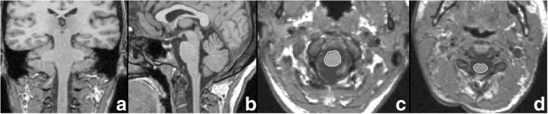
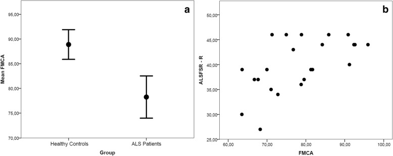
Spinal cord atrophy is one of the hallmarks of amyotrophic lateral sclerosis (ALS); however, it is not routinely assessed in routine clinical practice. In the present study, we evaluated whether spinal cord cross-sectional area measured at the foramen magnum level using a magnetic resonance imaging head scan represents a clinically meaningful measure to be added to the whole-brain volume assessment. Using an active surface approach, we measured the cord area at the foramen magnum and brain parenchymal fraction on T1-weighted three-dimensional spoiled gradient recalled head scans in two groups of subjects: 23 patients with ALS (males/females, 13/10; mean ± standard deviation [SD] age 61.7 ± 10.3 years; median ALS Functional Rating Scale-Revised score 39, range 27-46) and 18 age- and sex-matched healthy volunteers (mean ± SD age 55.7 ± 10.2 years). Spinal cord area at the foramen magnum was significantly less in patients than in control subjects and was significantly correlated with disability as measured with the ALS Functional Rating Scale-Revised (ρ = 0.593,  <  0.005). This correlation remained significant after taking into account inter-individual differences in brain parenchymal fraction (ρ = 0.684,  <  0.001). Our data show that spinal cord area at the foramen magnum correlates with disability in ALS independently of whole-brain atrophy, thus indicating its potential as a disease biomarker.

摘要

脊髓萎缩是肌萎缩侧索硬化症(ALS)的标志性特征之一;然而,在常规临床实践中并未对其进行常规评估。在本研究中,我们评估了使用磁共振成像头部扫描在枕骨大孔水平测量的脊髓横截面积是否代表一种有临床意义的测量方法,可添加到全脑体积评估中。我们采用主动表面法,在两组受试者的T1加权三维扰相梯度回波头部扫描上测量了枕骨大孔处的脊髓面积和脑实质分数:23例ALS患者(男/女,13/10;平均±标准差[SD]年龄61.7±10.3岁;ALS功能评定量表修订版评分中位数39,范围27 - 46)和18名年龄及性别匹配的健康志愿者(平均±SD年龄55.7±10.2岁)。患者枕骨大孔处的脊髓面积显著小于对照组,且与ALS功能评定量表修订版所测量的残疾程度显著相关(ρ = 0.593,< 0.005)。在考虑个体间脑实质分数差异后,这种相关性仍然显著(ρ = 0.684,< 0.001)。我们的数据表明,枕骨大孔处的脊髓面积与ALS患者的残疾程度相关,且独立于全脑萎缩,因此表明其作为疾病生物标志物的潜力。

https://cdn.ncbi.nlm.nih.gov/pmc/blobs/171e/6091748/9ea736ea3a62/41747_2018_45_Fig2_HTML.jpg
https://cdn.ncbi.nlm.nih.gov/pmc/blobs/171e/6091748/01fe951a9aed/41747_2018_45_Fig1_HTML.jpg
https://cdn.ncbi.nlm.nih.gov/pmc/blobs/171e/6091748/9ea736ea3a62/41747_2018_45_Fig2_HTML.jpg
https://cdn.ncbi.nlm.nih.gov/pmc/blobs/171e/6091748/01fe951a9aed/41747_2018_45_Fig1_HTML.jpg
https://cdn.ncbi.nlm.nih.gov/pmc/blobs/171e/6091748/9ea736ea3a62/41747_2018_45_Fig2_HTML.jpg

相似文献

1
Cord cross-sectional area at foramen magnum as a correlate of disability in amyotrophic lateral sclerosis.枕骨大孔处脊髓横截面积与肌萎缩侧索硬化症残疾程度的相关性
Eur Radiol Exp. 2018 Jun 22;2:13. doi: 10.1186/s41747-018-0045-6. eCollection 2018 Dec.
2
Association between brain and upper cervical spinal cord atrophy assessed by MRI and disease aggressiveness in amyotrophic lateral sclerosis.通过磁共振成像评估的脑和颈上段脊髓萎缩与肌萎缩侧索硬化症疾病侵袭性之间的关联
Neuroradiology. 2023 Sep;65(9):1395-1403. doi: 10.1007/s00234-023-03191-0. Epub 2023 Jul 17.
3
Diffusion anisotropy of the cervical cord is strictly associated with disability in amyotrophic lateral sclerosis.颈髓扩散各向异性与肌萎缩侧索硬化症的残疾程度密切相关。
J Neurol Neurosurg Psychiatry. 2007 May;78(5):480-4. doi: 10.1136/jnnp.2006.100032. Epub 2006 Oct 9.
4
Spinal cord atrophy correlates with disease duration and severity in amyotrophic lateral sclerosis.脊髓萎缩与肌萎缩侧索硬化症的疾病持续时间和严重程度相关。
Amyotroph Lateral Scler Frontotemporal Degener. 2014 Mar;15(1-2):93-7. doi: 10.3109/21678421.2013.852589. Epub 2013 Nov 13.
5
The upper cervical spinal cord in ALS assessed by cross-sectional and longitudinal 3T MRI.ALS 患者的上颈髓的横断面和纵向 3T MRI 评估。
Sci Rep. 2020 Feb 4;10(1):1783. doi: 10.1038/s41598-020-58687-z.
6
Brain and cord myelin water imaging: a progressive multiple sclerosis biomarker.脑和脊髓髓鞘水成像:一种进展性多发性硬化症生物标志物。
Neuroimage Clin. 2015 Oct 3;9:574-80. doi: 10.1016/j.nicl.2015.10.002. eCollection 2015.
7
A longitudinal diffusion tensor MRI study of the cervical cord and brain in amyotrophic lateral sclerosis patients.肌萎缩侧索硬化症患者颈髓和脑部的纵向扩散张量磁共振成像研究
J Neurol Neurosurg Psychiatry. 2009 Jan;80(1):53-5. doi: 10.1136/jnnp.2008.154252. Epub 2008 Oct 17.
8
Evidence for early neurodegeneration in the cervical cord of patients with primary progressive multiple sclerosis.原发性进行性多发性硬化症患者颈髓早期神经退行性变的证据。
Brain. 2015 Jun;138(Pt 6):1568-82. doi: 10.1093/brain/awv086. Epub 2015 Apr 10.
9
Diffusion tensor imaging reveals regional differences in the cervical spinal cord in amyotrophic lateral sclerosis.弥散张量成像显示肌萎缩侧索硬化症颈椎脊髓的区域性差异。
Neuroimage. 2010 Nov 1;53(2):576-83. doi: 10.1016/j.neuroimage.2010.06.060. Epub 2010 Jun 28.
10
Brain functional connectome abnormalities in amyotrophic lateral sclerosis are associated with disability and cortical hyperexcitability.肌萎缩侧索硬化症的脑功能连接组异常与残疾和皮质过度兴奋性有关。
Eur J Neurol. 2017 Dec;24(12):1507-1517. doi: 10.1111/ene.13461. Epub 2017 Oct 7.

引用本文的文献

1
Cervical spinal cord MRI in ALS individuals: a systematic review and meta-analysis.肌萎缩侧索硬化症患者的颈段脊髓磁共振成像:一项系统评价和荟萃分析。
Neuroimage Clin. 2025 Jun 26;47:103832. doi: 10.1016/j.nicl.2025.103832.
2
Real-time Quantification of in vivo cerebrospinal fluid velocity using fMRI inflow effect.利用功能磁共振成像流入效应实时定量测定体内脑脊液流速
bioRxiv. 2023 Nov 5:2023.08.14.553250. doi: 10.1101/2023.08.14.553250.
3
Neuroimaging findings in preclinical amyotrophic lateral sclerosis models-How well do they mimic the clinical phenotype? A systematic review.

本文引用的文献

1
Amyotrophic lateral sclerosis.肌萎缩性侧索硬化症。
Lancet. 2017 Nov 4;390(10107):2084-2098. doi: 10.1016/S0140-6736(17)31287-4. Epub 2017 May 25.
2
Neuroimaging Endpoints in Amyotrophic Lateral Sclerosis.肌萎缩侧索硬化症的神经影像学终点
Neurotherapeutics. 2017 Jan;14(1):11-23. doi: 10.1007/s13311-016-0484-9.
3
The value of magnetic resonance imaging as a biomarker for amyotrophic lateral sclerosis: a systematic review.磁共振成像作为肌萎缩侧索硬化症生物标志物的价值:一项系统综述。
临床前肌萎缩侧索硬化模型的神经影像学研究结果——它们对临床表型的模拟效果如何?一项系统综述。
Front Vet Sci. 2023 May 2;10:1135282. doi: 10.3389/fvets.2023.1135282. eCollection 2023.
4
Structural magnetic resonance imaging findings and histopathological correlations in motor neuron diseases-A systematic review and meta-analysis.运动神经元疾病的结构磁共振成像结果与组织病理学相关性——一项系统评价和荟萃分析
Front Neurol. 2022 Aug 30;13:947347. doi: 10.3389/fneur.2022.947347. eCollection 2022.
5
Spinal Cord Imaging in Amyotrophic Lateral Sclerosis: Historical Concepts-Novel Techniques.肌萎缩侧索硬化症的脊髓成像:历史概念与新技术
Front Neurol. 2019 Apr 12;10:350. doi: 10.3389/fneur.2019.00350. eCollection 2019.
BMC Neurol. 2016 Aug 27;16(1):155. doi: 10.1186/s12883-016-0672-6.
4
Cervical Spinal Cord Atrophy Profile in Adult SMN1-Linked SMA.成人SMN1相关脊髓性肌萎缩症的颈髓萎缩概况
PLoS One. 2016 Apr 18;11(4):e0152439. doi: 10.1371/journal.pone.0152439. eCollection 2016.
5
Cervical cord area measurement using volumetric brain magnetic resonance imaging in multiple sclerosis.多发性硬化症中使用容积性脑磁共振成像测量颈髓区域
Mult Scler Relat Disord. 2015 Jan;4(1):52-7. doi: 10.1016/j.msard.2014.11.004. Epub 2014 Nov 18.
6
Multi-parametric spinal cord MRI as potential progression marker in amyotrophic lateral sclerosis.多参数脊髓磁共振成像作为肌萎缩侧索硬化症潜在的病情进展标志物
PLoS One. 2014 Apr 22;9(4):e95516. doi: 10.1371/journal.pone.0095516. eCollection 2014.
7
Spinal cord atrophy correlates with disease duration and severity in amyotrophic lateral sclerosis.脊髓萎缩与肌萎缩侧索硬化症的疾病持续时间和严重程度相关。
Amyotroph Lateral Scler Frontotemporal Degener. 2014 Mar;15(1-2):93-7. doi: 10.3109/21678421.2013.852589. Epub 2013 Nov 13.
8
Method for simultaneous voxel-based morphometry of the brain and cervical spinal cord area measurements using 3D-MDEFT.使用 3D-MDEFT 进行基于体素的脑和颈椎脊髓区域测量的同步形态测量方法。
J Magn Reson Imaging. 2010 Nov;32(5):1242-7. doi: 10.1002/jmri.22340.
9
Cervical cord and brain grey matter atrophy independently associate with long-term MS disability.颈髓和脑灰质萎缩分别与多发性硬化症的长期残疾相关。
J Neurol Neurosurg Psychiatry. 2011 Apr;82(4):471-2. doi: 10.1136/jnnp.2010.205021. Epub 2010 Aug 14.
10
Reducing the impact of white matter lesions on automated measures of brain gray and white matter volumes.降低脑白质病变对脑灰白质体积自动测量的影响。
J Magn Reson Imaging. 2010 Jul;32(1):223-8. doi: 10.1002/jmri.22214.