University of Liverpool, Liverpool, UK.
University of Regensburg, Regensburg, Germany.
Neuroimage Clin. 2018 Jun 25;20:1-6. doi: 10.1016/j.nicl.2018.06.027. eCollection 2018.
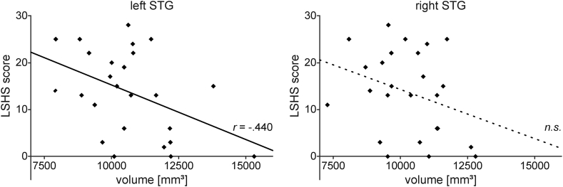
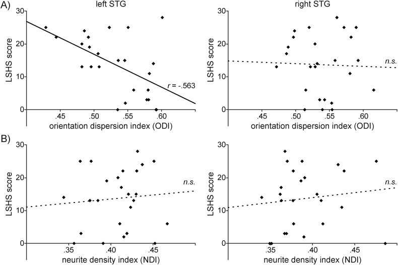
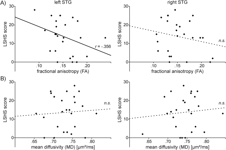
Previous studies reported that the volume of the left superior temporal gyrus (STG) is reduced in patients with schizophrenia and negatively correlated with hallucination severity. Moreover, diffusion-tensor imaging studies suggested a relationship between the brain microstructure in the STG of patients and auditory hallucinations. Hallucinations are also experienced in non-patient groups. This study investigated the relationship between hallucination proneness and the brain structure of the STG. Hallucination proneness was assessed by the Launey Slade Hallucination Scale (LSHS) in 25 healthy individuals who varied in their propensity to hear voices. Brain volume and microstructure of the STG was assessed by magnetic resonance imaging (MRI). Microstructure was examined by conventional diffusion-tensor imaging as well as by neurite orientation dispersion and density imaging (NODDI). The latter decomposes diffusion-based MRI into multiple compartments that characterize the brain microstructure by its neurite complexity known as orientation dispersion (ODI) and by its neurite density (NDI). Hallucination proneness was negatively correlated with the volume and microstructure (fractional anisotropy, neurite complexity) of the left but not the right STG. The strongest relationship ( = -0.563) was observed for neurite complexity (ODI). No correlation was observed for neurite density (NDI). These findings suggest that there is a relationship between the volume and the microstructure of the left STG and hallucination proneness. Dendritic complexity (but not neurite density) is inversely related to hallucination proneness. Metrics based on multi-compartment diffusion models seem to be more sensitive for hallucination-related neural processes than conventional MRI-based metrics.
先前的研究报告指出,精神分裂症患者的左侧颞上回(STG)体积减小,且与幻觉严重程度呈负相关。此外,弥散张量成像研究表明,患者 STG 的大脑微观结构与听觉幻觉之间存在关系。非患者群体也会经历幻觉。本研究旨在探讨易出现幻觉与 STG 大脑结构之间的关系。采用 Launey Slade 幻觉量表(LSHS)评估 25 名健康个体的幻觉易感性,这些个体的听觉幻觉倾向存在差异。采用磁共振成像(MRI)评估 STG 的脑体积和微观结构。通过常规弥散张量成像以及神经丝取向分散度和密度成像(NODDI)检查微观结构。后者将基于扩散的 MRI 分解为多个隔室,通过其神经丝复杂度(称为取向分散度(ODI))和神经丝密度(NDI)来描述大脑微观结构。幻觉易感性与左侧而非右侧 STG 的体积和微观结构(各向异性分数、神经丝复杂度)呈负相关。最强的相关性(r= -0.563)是在神经丝复杂度(ODI)中观察到的。神经丝密度(NDI)没有相关性。这些发现表明,左侧 STG 的体积和微观结构与幻觉易感性之间存在关系。树突复杂性(而非神经丝密度)与幻觉易感性呈负相关。基于多隔室扩散模型的指标似乎比基于常规 MRI 的指标更能敏感地反映与幻觉相关的神经过程。