Kyrölä Kati Kristiina, Salme Järvenpää, Tuija Järviluoma, Tero Irmola, Eero Kauppinen, Arja Häkkinen
Department of Orthopedics and Traumatology, Central Hospital of Central Hospital, Jyväskylä, Finland.
Department of Physical Medicine and Rehabilitation, Central Hospital of Central Hospital, Jyväskylä, Finland.
Neurospine. 2018 Jun;15(2):175-181. doi: 10.14245/ns.1836054.027. Epub 2018 Jun 19.
To evaluate the intra- and interrater reliability (I-IR) of sagittal spinopelvic parameters from digital full-spine plain radiographs with basic software tools in an unselected adult population with degenerative spinal complaints who were evaluated for surgery.
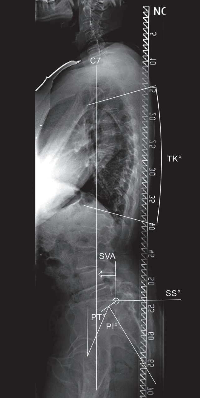
Forty-nine adult full-spine digital radiographs were measured twice by 3 independent observers, including an experienced spine surgeon, an experienced radiologist, and a resident orthopedic surgeon. Clinical picture archiving and communication system workstations and software tools were used and landmarks were set manually. The I-IR of the sagittal vertical axis (SVA), pelvic tilt (PT), pelvic incidence (PI), sacral slope (SS), and thoracic kyphosis in T4-T12 (TK) were assessed.
The intrarater intraclass correlation coefficient (ICC) scores varied from 0.82 to 0.99. The interrater ICC scores ranged from 0.78 to 0.99. The intrarater standard error of measurement (SEM) values for SS, PT, PI, and TK varied from 0.8° to 5.0°, and the interrater SEM values ranged from 2.5° to 6.2°, depending on the parameter and the reading round. The I-IR SEM values for SVA varied from 2.2 to 5.7 mm and from 4.6 to 5.0 mm, respectively. Kappa values were >0.88 for all readers. The intrarater variability was the smallest for the most experienced rater.
The I-IR of measuring sagittal spinopelvic parameters on digital full-spine images with basic software tools was high. Parameters consisting of several anatomic landmarks were more liable to error. Rater experience had a positive influence on reliability and repeatability. Reader experience should be assessed before accepting measurements for surgical planning and the interpretation of surgical correction during postoperative follow-up.
使用基本软件工具,对未经挑选的患有退行性脊柱疾病且接受手术评估的成年人群的数字化全脊柱平片矢状位脊柱骨盆参数的组内和组间可靠性(I-IR)进行评估。
49例成人全脊柱数字化X线片由3名独立观察者测量两次,包括一名经验丰富的脊柱外科医生、一名经验丰富的放射科医生和一名骨科住院医师。使用临床图像存档与通信系统工作站及软件工具,并手动设置标志点。评估矢状垂直轴(SVA)、骨盆倾斜度(PT)、骨盆入射角(PI)、骶骨倾斜度(SS)以及T4-T12节段胸椎后凸(TK)的I-IR。
组内类内相关系数(ICC)评分在0.82至0.99之间。组间ICC评分在0.78至0.99之间。SS、PT、PI和TK的组内测量标准误(SEM)值在0.8°至5.0°之间,组间SEM值在2.5°至6.2°之间,具体取决于参数和测量轮次。SVA的I-IR SEM值分别在2.2至5.7毫米和4.6至5.0毫米之间。所有阅片者的kappa值均>0.88。对于经验最丰富的阅片者,组内变异性最小。
使用基本软件工具在数字化全脊柱图像上测量矢状位脊柱骨盆参数的I-IR较高。由多个解剖标志点组成的参数更容易出现误差。阅片者经验对可靠性和可重复性有积极影响。在接受测量结果用于手术规划以及术后随访期间手术矫正的解读之前,应评估阅片者经验。