• 文献检索
  • 文档翻译
  • 深度研究
  • 学术资讯
  • Suppr Zotero 插件Zotero 插件
  • 邀请有礼
  • 套餐&价格
  • 历史记录
应用&插件
Suppr Zotero 插件Zotero 插件浏览器插件Mac 客户端Windows 客户端微信小程序
定价
高级版会员购买积分包购买API积分包
服务
文献检索文档翻译深度研究API 文档MCP 服务
关于我们
关于 Suppr公司介绍联系我们用户协议隐私条款
关注我们

Suppr 超能文献

核心技术专利:CN118964589B侵权必究
粤ICP备2023148730 号-1Suppr @ 2026

文献检索

告别复杂PubMed语法,用中文像聊天一样搜索,搜遍4000万医学文献。AI智能推荐,让科研检索更轻松。

立即免费搜索

文件翻译

保留排版,准确专业,支持PDF/Word/PPT等文件格式,支持 12+语言互译。

免费翻译文档

深度研究

AI帮你快速写综述,25分钟生成高质量综述,智能提取关键信息,辅助科研写作。

立即免费体验

用于人工智能准确性分析的临床基准数据集:定量分析骨盆倾斜的放射影像标注。

Clinical benchmark dataset for AI accuracy analysis: quantifying radiographic annotation of pelvic tilt.

机构信息

Sydney Musculoskeletal Health and The Kolling Institute, Northern Clinical School, Faculty of Medicine and Health and the Northern Sydney Local Health District, Sydney, NSW, Australia.

University Hospitals Leuven, Department of Orthopedic Surgery, Leuven, Belgium.

出版信息

Sci Data. 2024 Oct 22;11(1):1162. doi: 10.1038/s41597-024-04003-7.

DOI:10.1038/s41597-024-04003-7
PMID:39438488
原文链接:https://pmc.ncbi.nlm.nih.gov/articles/PMC11496730/
Abstract

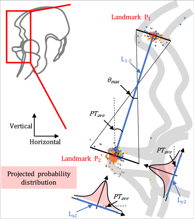
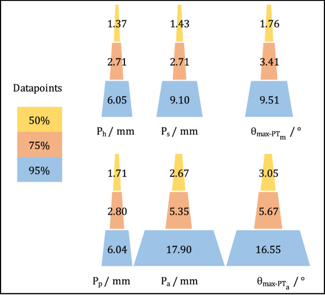
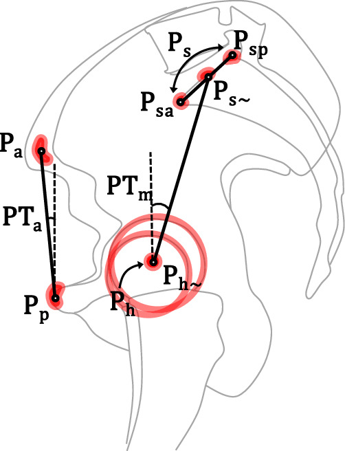
Radiographic landmark annotation determines patients' anatomical parameters and influences diagnoses. However, challenges arise from ambiguous region-based definitions, human error, and image quality variations, potentially compromising patient care. Additionally, AI landmark localization often presents its predictions in a probability-based heatmap format, which lacks a corresponding clinical standard for accuracy validation. This Data Descriptor presents a clinical benchmark dataset for pelvic tilt landmarks, gathered through a probabilistic approach to measure annotation accuracy within clinical environments. A retrospective analysis of 115 pelvic sagittal radiographs was conducted for annotating pelvic tilt parameters by five annotators, revealing landmark cloud sizes of 6.04 mm-17.90 mm at a 95% dataset threshold, corresponding to 9.51°-16.55° maximum angular disagreement in clinical settings. The outcome provides a quantified point cloud dataset for each landmark corresponding to different probabilities, which enables assessment of directional annotation distribution and parameter-wise impact, providing clinical benchmarks. The data is readily reusable for AI studies analyzing the same landmarks, and the method can be easily replicated for establishing clinical accuracy benchmarks of other landmarks.

摘要

影像学标志点标注决定了患者的解剖参数,并影响诊断。然而,基于区域的定义不明确、人为误差和图像质量变化等挑战,可能会影响患者的治疗。此外,人工智能标志点定位通常以基于概率的热图格式呈现其预测结果,这缺乏相应的临床准确性验证标准。本数据描述符提供了一个用于骨盆倾斜标志点的临床基准数据集,该数据集是通过一种概率方法在临床环境中测量标注准确性而收集的。通过对 115 张骨盆矢状面射线照片进行回顾性分析,由五名标注者对骨盆倾斜参数进行标注,在 95%数据集阈值下,标志点云大小为 6.04mm-17.90mm,这对应于临床环境下最大角度差异 9.51°-16.55°。该结果为每个标志点提供了对应不同概率的量化点云数据集,可用于评估定向标注分布和参数影响,为临床提供基准。该数据可方便地重复用于分析相同标志点的人工智能研究,并且可以轻松复制该方法以建立其他标志点的临床准确性基准。

https://cdn.ncbi.nlm.nih.gov/pmc/blobs/0be5/11496730/f5f7ccef4281/41597_2024_4003_Fig4_HTML.jpg
https://cdn.ncbi.nlm.nih.gov/pmc/blobs/0be5/11496730/00f840e31523/41597_2024_4003_Fig1_HTML.jpg
https://cdn.ncbi.nlm.nih.gov/pmc/blobs/0be5/11496730/87d627b22638/41597_2024_4003_Fig2_HTML.jpg
https://cdn.ncbi.nlm.nih.gov/pmc/blobs/0be5/11496730/356f3f6cb1a5/41597_2024_4003_Fig3_HTML.jpg
https://cdn.ncbi.nlm.nih.gov/pmc/blobs/0be5/11496730/f5f7ccef4281/41597_2024_4003_Fig4_HTML.jpg

相似文献

1
Clinical benchmark dataset for AI accuracy analysis: quantifying radiographic annotation of pelvic tilt.用于人工智能准确性分析的临床基准数据集:定量分析骨盆倾斜的放射影像标注。
Sci Data. 2024 Oct 22;11(1):1162. doi: 10.1038/s41597-024-04003-7.
2
Inadequate Annotation and Its Impact on Pelvic Tilt Measurement in Clinical Practice.标注不充分及其对临床实践中骨盆倾斜度测量的影响。
J Clin Med. 2024 Feb 28;13(5):1394. doi: 10.3390/jcm13051394.
3
The Sacro-femoral-pubic Angle Is Unreliable to Estimate Pelvic Tilt: A Meta-analysis.骶耻-耻骨角估计骨盆倾斜不可靠:一项荟萃分析。
Clin Orthop Relat Res. 2023 Oct 1;481(10):1928-1936. doi: 10.1097/CORR.0000000000002650. Epub 2023 Apr 18.
4
A Modeling Study of a Patient-specific Safe Zone for THA: Calculation, Validation, and Key Factors Based on Standing and Sitting Sagittal Pelvic Tilt.基于站立和坐位矢状位骨盆倾斜的全髋关节置换术患者安全区的建模研究:计算、验证及关键因素
Clin Orthop Relat Res. 2022 Jan 1;480(1):191-205. doi: 10.1097/CORR.0000000000001923.
5
Does Compensatory Anterior Pelvic Tilt Decrease After Bilateral Periacetabular Osteotomy?双侧髋臼周围截骨术后补偿性前骨盆倾斜是否减少?
Clin Orthop Relat Res. 2019 May;477(5):1168-1175. doi: 10.1097/CORR.0000000000000560.
6
Radiographic analysis of the sagittal alignment and balance of the spine in asymptomatic subjects.无症状受试者脊柱矢状位排列及平衡的影像学分析。
J Bone Joint Surg Am. 2005 Feb;87(2):260-7. doi: 10.2106/JBJS.D.02043.
7
Small Random Angular Variations in Pelvic Tilt and Lower Extremity Can Cause Error in Static Image-based Preoperative Hip Arthroplasty Planning: A Computer Modeling Study.骨盆倾斜和下肢的微小随机角度变化会导致基于静态图像的术前髋关节置换术规划出现误差:一项计算机建模研究。
Clin Orthop Relat Res. 2022 Apr 1;480(4):818-828. doi: 10.1097/CORR.0000000000002106.
8
Auxiliary diagnosis of developmental dysplasia of the hip by automated detection of Sharp's angle on standardized anteroposterior pelvic radiographs.通过自动检测标准化骨盆前后位X线片上的夏普角辅助诊断发育性髋关节发育不良。
Medicine (Baltimore). 2019 Dec;98(52):e18500. doi: 10.1097/MD.0000000000018500.
9
Validation of a new computer-assisted tool to measure spino-pelvic parameters.一种用于测量脊柱-骨盆参数的新型计算机辅助工具的验证
Spine J. 2015 Dec 1;15(12):2493-502. doi: 10.1016/j.spinee.2015.08.067. Epub 2015 Sep 4.
10
Development and validation of an artificial intelligence model to accurately predict spinopelvic parameters.开发和验证一种人工智能模型,以准确预测脊柱骨盆参数。
J Neurosurg Spine. 2024 Mar 29;41(1):88-96. doi: 10.3171/2024.1.SPINE231252. Print 2024 Jul 1.

引用本文的文献

1
Evaluating Alternative Registration Planes in Imageless, Computer-Assisted Navigation Systems for Direct Anterior Total Hip Arthroplasty.评估无影像、计算机辅助导航系统中直接前侧全髋关节置换术的替代注册平面。
Sensors (Basel). 2024 Nov 4;24(21):7092. doi: 10.3390/s24217092.

本文引用的文献

1
Advances in imaging for pre-surgical planning in hip resurfacing arthroplasty.髋关节表面置换术中术前规划成像技术的进展。
Orthop Traumatol Surg Res. 2024 Oct;110(6):103908. doi: 10.1016/j.otsr.2024.103908. Epub 2024 May 19.
2
Inadequate Annotation and Its Impact on Pelvic Tilt Measurement in Clinical Practice.标注不充分及其对临床实践中骨盆倾斜度测量的影响。
J Clin Med. 2024 Feb 28;13(5):1394. doi: 10.3390/jcm13051394.
3
Letter to the Editor Regarding the Article "Comparison of Transfer Learning Models in Pelvic Tilt and Rotation Measurement in Pediatric Anteroposterior Pelvic Radiographs".
致编辑的信:关于文章《儿科前后位骨盆X线片中骨盆倾斜和旋转测量的迁移学习模型比较》
J Imaging Inform Med. 2024 Jun;37(3):1259-1260. doi: 10.1007/s10278-024-01025-w. Epub 2024 Feb 9.
4
Evaluating Pelvic Tilt Using the Pelvic Antero-Posterior Projection Images: A Systematic Review.使用骨盆前后位投影图像评估骨盆倾斜:一项系统评价。
J Arthroplasty. 2024 Apr;39(4):1108-1116.e2. doi: 10.1016/j.arth.2023.10.035. Epub 2023 Oct 21.
5
The Sacro-femoral-pubic Angle Is Unreliable to Estimate Pelvic Tilt: A Meta-analysis.骶耻-耻骨角估计骨盆倾斜不可靠:一项荟萃分析。
Clin Orthop Relat Res. 2023 Oct 1;481(10):1928-1936. doi: 10.1097/CORR.0000000000002650. Epub 2023 Apr 18.
6
Correlations Analysis of Different Pelvic Tilt Definitions: A Preliminary Study.不同骨盆倾斜度定义的相关性分析:一项初步研究。
HSS J. 2023 May;19(2):187-192. doi: 10.1177/15563316221136128. Epub 2022 Nov 26.
7
Automatic recognition of whole-spine sagittal alignment and curvature analysis through a deep learning technique.通过深度学习技术实现全脊柱矢状位排列和曲率的自动识别。
Eur Spine J. 2022 Aug;31(8):2092-2103. doi: 10.1007/s00586-022-07189-9. Epub 2022 Apr 2.
8
Deep learning-enabled coronary CT angiography for plaque and stenosis quantification and cardiac risk prediction: an international multicentre study.深度学习辅助冠状动脉 CT 血管造影术进行斑块和狭窄定量及心脏风险预测:一项国际多中心研究。
Lancet Digit Health. 2022 Apr;4(4):e256-e265. doi: 10.1016/S2589-7500(22)00022-X.
9
Spinopelvic measurements of sagittal balance with deep learning: systematic review and critical evaluation.深度学习的矢状位平衡的脊柱骨盆测量:系统综述和批判性评估。
Eur Spine J. 2022 Aug;31(8):2031-2045. doi: 10.1007/s00586-022-07155-5. Epub 2022 Mar 12.
10
2-step deep learning model for landmarks localization in spine radiographs.两步深度学习模型用于脊柱 X 光片中的地标定位。
Sci Rep. 2021 May 4;11(1):9482. doi: 10.1038/s41598-021-89102-w.