Allergy Clinic, Copenhagen University Hospital Gentofte, Copenhagen, Denmark.
Medical Prognosis Institute, Hoersholm, Denmark.
Immun Inflamm Dis. 2018 Dec;6(4):416-427. doi: 10.1002/iid3.226. Epub 2018 Jul 10.
Mast cells are the primary effector cells of allergy. This study aimed at characterizing human peripheral blood-derived mast cells (PBdMC) from peanut allergic and non-allergic subjects by investigating whether the molecular and stimulus-response profile of PBdMC discriminate between peanut allergic and healthy individuals.
PBdMC were generated from eight peanut allergic and 10 non-allergic subjects. The molecular profile (cell surface receptor expression) was assessed using flow cytometry. The stimulus-response profile (histamine release induced by secretagogues, secretion of cytokines/chemokines and changes in miRNA expression following anti-IgE activation) was carried out with histamine release test, luminex multiplex assay and miRNA arrays.
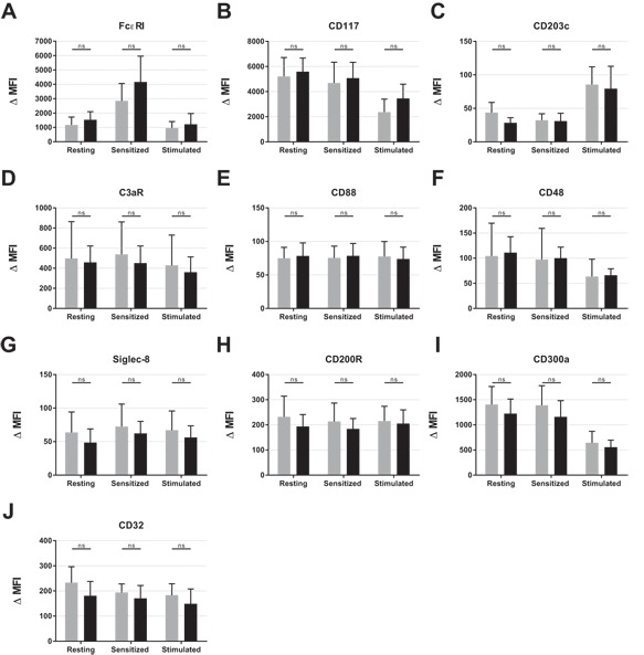
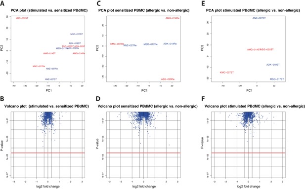
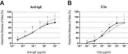
Expression of activating receptors (FcϵRI, CD48, CD88, CD117, and C3aR) on PBdMC was not different among peanut allergic and non-allergic subjects. Likewise, inhibitory receptors (CD32, CD200R, CD300a, and siglec-8) displayed comparable levels of expression. Both groups of PBdMC were unresponsive to substance P, compound 48/80 and C5a but released comparable levels of histamine when stimulated with anti-IgE and C3a. Interestingly, among the secreted cytokines/chemokines (IL-8, IL-10, IL-13, IL-23, IL-31, IL-37, MCP-1, VEGF, GM-CSF) PBdMC from peanut allergic subjects showed a different secretion pattern of IL-31 compared to non-allergic subjects. Investigating miRNA expression from resting or activated PBdMC revealed no significantly difference between peanut allergic and non-allergic subjects.
The molecular and stimulus-response profile revealed that PBdMC from peanut allergic subjects differently express IL-31 compared to non-allergic subjects. However, since only one altered parameter was found among 893 investigated, it is still questionable if the pathophysiological mechanisms of peanut allergy are revealed in PBdMC.
肥大细胞是过敏的主要效应细胞。本研究旨在通过研究外周血来源的肥大细胞(PBdMC)的分子和刺激反应特征是否可以区分花生过敏和健康个体,从而对来自花生过敏和非过敏个体的 PBdMC 进行特征描述。
从 8 名花生过敏和 10 名非过敏个体中生成 PBdMC。使用流式细胞术评估分子谱(细胞表面受体表达)。通过组胺释放试验、Luminex 多重分析和 miRNA 阵列进行刺激反应谱(由 secretagogues 诱导的组胺释放、细胞因子/趋化因子的分泌以及抗 IgE 激活后 miRNA 表达的变化)。
花生过敏和非过敏个体的 PBdMC 上激活受体(FcεRI、CD48、CD88、CD117 和 C3aR)的表达无差异。同样,抑制性受体(CD32、CD200R、CD300a 和 siglec-8)也表现出相似的表达水平。两组 PBdMC 对 P 物质、化合物 48/80 和 C5a 均无反应,但用抗 IgE 和 C3a 刺激时释放的组胺水平相当。有趣的是,在分泌的细胞因子/趋化因子(IL-8、IL-10、IL-13、IL-23、IL-31、IL-37、MCP-1、VEGF、GM-CSF)中,与非过敏个体相比,花生过敏个体的 PBdMC 表现出不同的 IL-31 分泌模式。从静止或激活的 PBdMC 中检测 miRNA 表达,花生过敏和非过敏个体之间没有发现显著差异。
分子和刺激反应谱表明,与非过敏个体相比,花生过敏个体的 PBdMC 表达不同的 IL-31。然而,由于在 893 个研究参数中仅发现一个改变的参数,因此仍然存在疑问,即花生过敏的病理生理机制是否在 PBdMC 中得到揭示。