Institute of Cancer and Translational Medicine, Department of Anatomy and Cell Biology, University of Oulu, FI-90014, Oulu, Finland.
Medical Research Center Oulu, FI-90014, Oulu, Finland.
Sci Rep. 2018 Jul 11;8(1):10457. doi: 10.1038/s41598-018-28699-x.
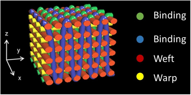
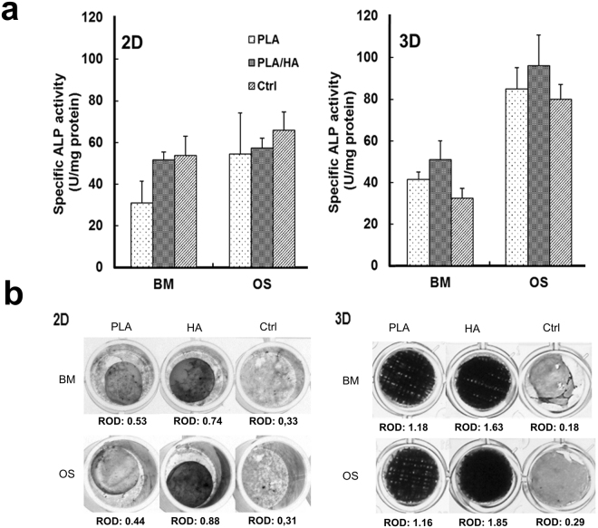
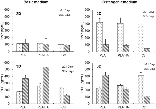
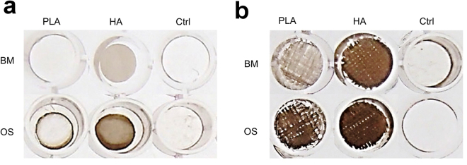
Fiber-based scaffolds produced by textile manufacturing technology offer versatile materials for tissue engineering applications since a wide range of crucial scaffold parameters, including porosity, pore size and interconnectivity, can be accurately controlled using 3D weaving. In this study, we developed a weavable, bioactive biodegradable composite fiber from poly (lactic acid) (PLA) and hydroxyapatite powder by melt spinning. Subsequently, scaffolds of these fibers were fabricated by 3D weaving. The differentiation of human mesenchymal stem cells (hMSCs) in vitro was studied on the 3D scaffolds and compared with differentiation on 2D substrates having the same material composition. Our data showed that the 3D woven scaffolds have a major impact on hMSCs proliferation and activation. The 3D architecture supports the differentiation of the hMSCs into osteoblast cells and enhances the production of mineralized bone matrix. The present study further confirms that a 3D scaffold promotes hMSCs differentiation into the osteoblast-lineage and bone mineralization.
基于纤维的支架由纺织制造技术生产,为组织工程应用提供了多种材料,因为使用 3D 编织可以精确控制包括孔隙率、孔径和连通性在内的广泛关键支架参数。在这项研究中,我们通过熔融纺丝开发了一种可编织的、具有生物活性的可生物降解的聚乳酸(PLA)和羟基磷灰石粉末复合材料纤维。随后,通过 3D 编织制造了这些纤维的支架。在体外研究了这些 3D 支架上的人骨髓间充质干细胞(hMSCs)的分化,并与具有相同材料组成的 2D 基质上的分化进行了比较。我们的数据表明,3D 编织支架对 hMSCs 的增殖和激活有重大影响。3D 结构支持 hMSCs 分化为成骨细胞,并增强矿化骨基质的产生。本研究进一步证实,3D 支架可促进 hMSCs 向成骨细胞系分化和骨矿化。