Centre International de Recherche en Infectiologie, Institut National de la Santé et de la Recherche Médicale, U1111, Centre National de la Recherche Scientifique, UMR5308, Université de Lyon 1, École Normale Supérieure de Lyon, Team "Pathogenesis of Staphylococcal Infections", Lyon, France.
Institut des Agents Infectieux, Hospices Civils de Lyon, Lyon, France.
Front Cell Infect Microbiol. 2018 Jun 28;8:221. doi: 10.3389/fcimb.2018.00221. eCollection 2018.
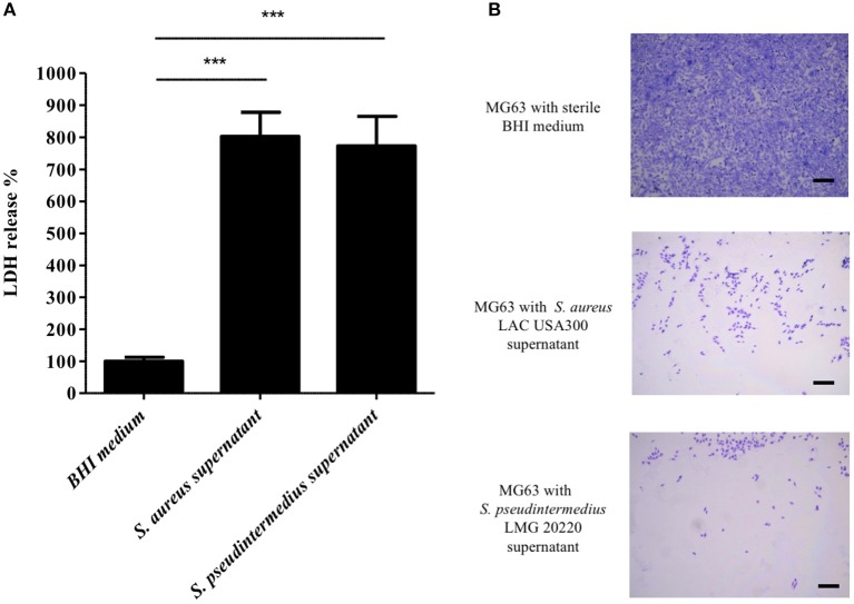
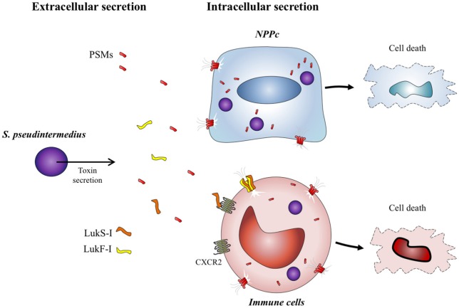
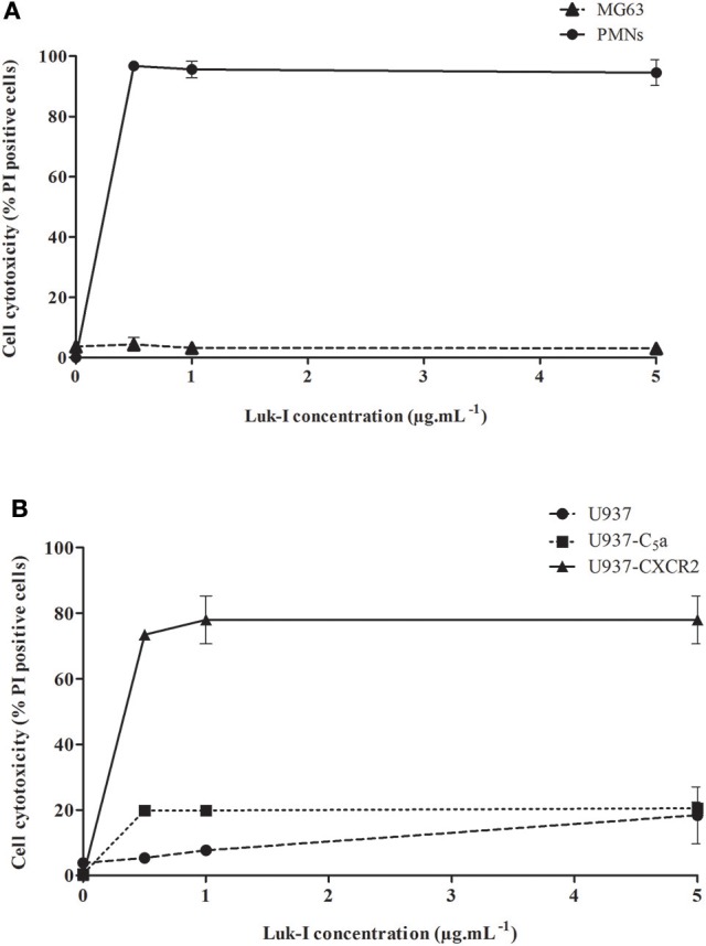
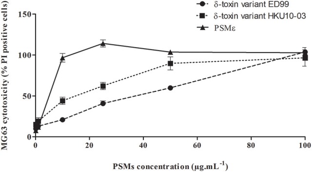
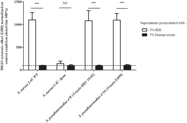
is responsible for severe and necrotizing infections in humans and dogs. Contrary to , the pathophysiological mechanisms involved in this virulence are incompletely understood. We previously showed the intracellular cytotoxicity induced after internalization of . Herein, we aimed to identify the virulence factors responsible for this cytotoxic activity. After addition of filtered supernatants in culture cell media, MG63 cells, used as representative of non-professional phagocytic cells (NPPc), released a high level of LDH, indicating that the cytotoxicity was mainly mediated by secreted factors. Accordingly, we focused our attention on toxins. analysis found the presence of two PSMs (δ-toxin and PSMε) as well as Luk-I leukotoxin, the presence of which was confirmed by PCR in all clinical strains tested ( = 17). Recombinant Luk-I leukotoxin had no cytotoxic activity on NPPc but the ectopic expression of the CXCR2 receptor in U937 cells conferred cytotoxity to Luk-I. This is in agreement with the lack of Luk-I effect on NPPc and the previous report of Luk-I cytoxic activity on immune cells. Contrary to Luk-I, synthetic δ-toxin and PSMε had a strong cytotoxic activity on NPPc. The secretion of δ-toxin and PSMε at cytotoxic concentrations by in culture supernatant was confirmed by HPLC-MS. In addition, the supplementation of such supernatants with human serum, known to inhibit PSM, induced a complete abolition of cytotoxicity which indicates that PSMs are the key players in the cytotoxic phenotype of NPPc. The results suggest that the severity of infections is, at least in part, explained by a combined action of Luk-I that specifically targets immune cells expressing the CXCR2 receptor, and PSMs that disrupt cell membranes whatever the cell types. The present study strengthens the key role of PSMs in virulence of the different species belonging to genus.
是人类和犬类严重坏死性感染的病原体。与不同,其毒力涉及的病理生理机制尚未完全阐明。我们之前已经证明了 内化后诱导的细胞内细胞毒性。在此,我们旨在确定导致这种细胞毒性的毒力因子。在添加过滤后的 上清液后,MG63 细胞作为非专业吞噬细胞 (NPPc) 的代表,释放出高水平的 LDH,表明细胞毒性主要由分泌因子介导。因此,我们将注意力集中在 毒素上。分析发现存在两种 PSM(δ-毒素和 PSMε)以及 Luk-I 白细胞毒素,所有测试的临床菌株(= 17)均通过 PCR 证实存在这些毒素。重组 Luk-I 白细胞毒素对 NPPc 没有细胞毒性,但在 U937 细胞中异位表达 CXCR2 受体赋予了 Luk-I 细胞毒性。这与 Luk-I 对 NPPc 没有影响以及 Luk-I 对免疫细胞的细胞毒性作用的先前报道一致。与 Luk-I 相反,合成的 δ-毒素和 PSMε 对 NPPc 具有很强的细胞毒性。通过 HPLC-MS 证实了 在培养上清液中以细胞毒性浓度分泌 δ-毒素和 PSMε。此外,在这些上清液中添加已知抑制 PSM 的人血清会导致细胞毒性完全消除,这表明 PSM 是 NPPc 细胞毒性表型的关键因素。这些结果表明, 感染的严重性至少部分是由 Luk-I 的共同作用解释的,Luk-I 特异性靶向表达 CXCR2 受体的免疫细胞,而 PSM 破坏细胞膜,无论细胞类型如何。本研究进一步证实了 PSM 在 属的不同种属的毒力中的关键作用。