Qian Neng, Li Xiaobo, Wang Xinhong, Wu Chungen, Yin Lianhua, Zhi Xiuling
School of Basic Medicine, Shanghai University of Medicine and Health Science, Shanghai 201318, P.R. China.
Department of Physiology and Pathophysiology, School of Basic Medical Sciences, Fudan University, Shanghai 200032, P.R. China.
Oncol Lett. 2018 Aug;16(2):1513-1520. doi: 10.3892/ol.2018.8856. Epub 2018 May 31.
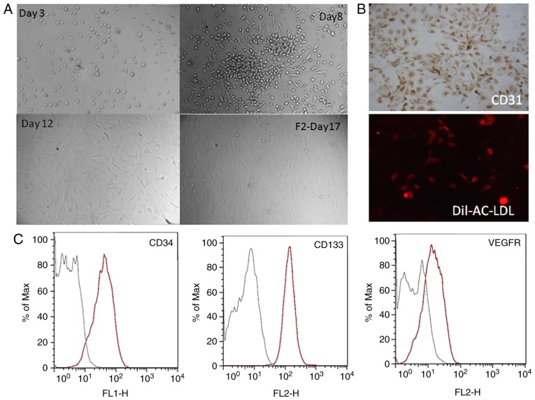
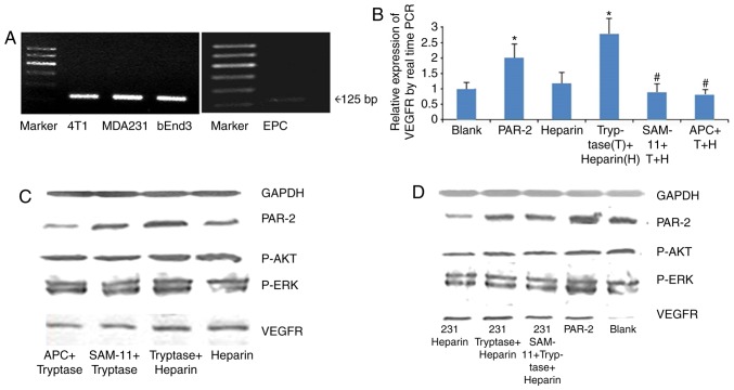
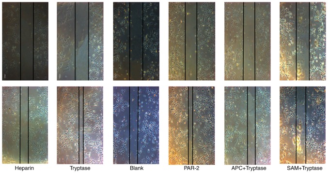
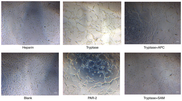
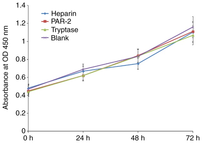
Mast cells have been demonstrated to accumulate around and within solid tumors of numerous types, and express a number of pro-angiogenic compounds, including tryptase. They may serve an early role in angiogenesis within developing tumors. In the present study, the role and mechanism of tryptase in the activation of endothelial progenitor cells (EPCs) in breast cancer angiogenesis were evaluated. Human umbilical cord blood EPCs were isolated and cultured. MB-MDA-231 breast cancer cells were then pretreated with tryptase, and the conditioned medium was collected. The effects of tryptase on the migratory and angiogenesis abilities of EPCs were determined using wound-healing and tube formation assays, respectively. The effect of tryptase on the proliferation of EPCs was detected using a Cell Counting Kit-8 assay. Alterations in proteinase activated receptor (PAR)-2, phosphorylated (p)-protein kinase B (AKT), p-extracellular signal-regulated kinase (p-ERK) and vascular endothelial growth factor receptor (VEGFR)-2 expression were analyzed, in tryptase or conditioned medium-treated EPCs, by western blot analysis and reverse transcription-quantitative polymerase chain reaction. It was confirmed that the EPCs expressed PAR-2; and that tryptase treatment promoted the migration and tube formation of EPCs. Treatment with a PAR-2 agonist had a similar effect to tryptase, whereas treatment with a tryptase inhibitor, APC366, or a PAR-2 inhibitor, SAM 11, inhibited the effect of tryptase treatment. Tryptase and PAR-2 agonists did not affect the rate of EPC proliferation. MB-MDA-231 cells also expressed PAR-2. Treatment with tryptase or conditioned medium increased the expression of PAR-2, p-AKT, p-ERK and VEGFR-2 in EPCs. In conclusion, tryptase activated EPCs via PAR-2-mediated AKT and ERK signaling pathway activation, thereby enhancing angiogenesis in breast cancer.
肥大细胞已被证明会在多种实体瘤周围及内部聚集,并表达多种促血管生成化合物,包括组织蛋白酶。它们可能在肿瘤发生发展过程中的血管生成中发挥早期作用。在本研究中,评估了组织蛋白酶在乳腺癌血管生成中激活内皮祖细胞(EPC)的作用及机制。分离并培养人脐带血EPC。然后用组织蛋白酶预处理MB-MDA-231乳腺癌细胞,并收集条件培养基。分别使用伤口愈合试验和管形成试验测定组织蛋白酶对EPC迁移和血管生成能力的影响。使用细胞计数试剂盒-8检测组织蛋白酶对EPC增殖的影响。通过蛋白质印迹分析和逆转录-定量聚合酶链反应分析组织蛋白酶或条件培养基处理的EPC中蛋白酶激活受体(PAR)-2、磷酸化(p)-蛋白激酶B(AKT)、p-细胞外信号调节激酶(p-ERK)和血管内皮生长因子受体(VEGFR)-2表达的变化。证实EPC表达PAR-2;并且组织蛋白酶处理促进了EPC的迁移和管形成。用PAR-2激动剂处理具有与组织蛋白酶相似的作用,而用组织蛋白酶抑制剂APC366或PAR-2抑制剂SAM 11处理则抑制了组织蛋白酶处理的效果。组织蛋白酶和PAR-2激动剂不影响EPC的增殖率。MB-MDA-231细胞也表达PAR-2。用组织蛋白酶或条件培养基处理增加了EPC中PAR-2、p-AKT、p-ERK和VEGFR-2的表达。总之,组织蛋白酶通过PAR-2介导的AKT和ERK信号通路激活来激活EPC,从而增强乳腺癌中的血管生成。