Wu Jian-Ching, Tsai Han-En, Liu Guei-Sheung, Wu Chieh-Shan, Tai Ming-Hong
1Doctoral Degree Program in Marine Biotechnology, National Sun Yat-sen University, 70 Lien-Hai Road, Kaohsiung, 80424 Taiwan.
2Doctoral Degree Program in Marine Biotechnology, Academia Sinica, 128 Academia Road, Section 2, Nankang, Taipei, 11529 Taiwan.
Cell Death Discov. 2018 Jul 10;4:11. doi: 10.1038/s41420-018-0070-5. eCollection 2018.
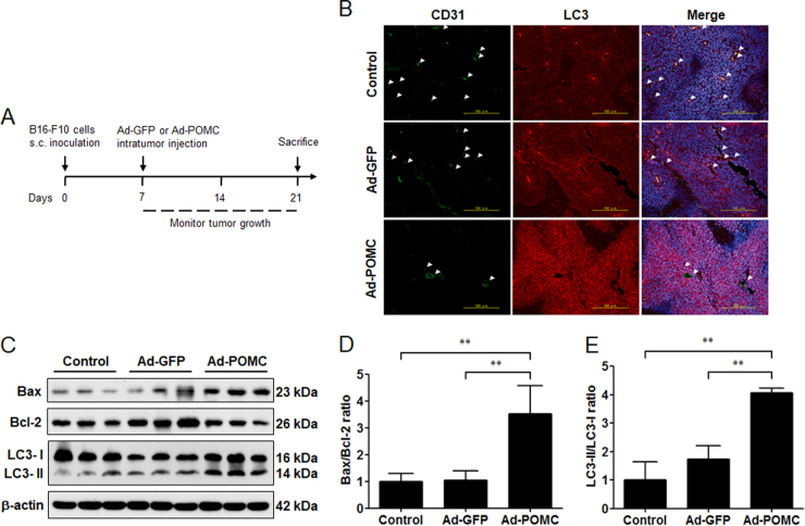
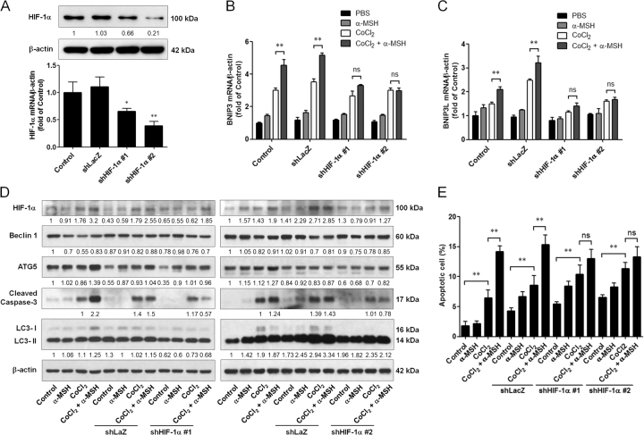
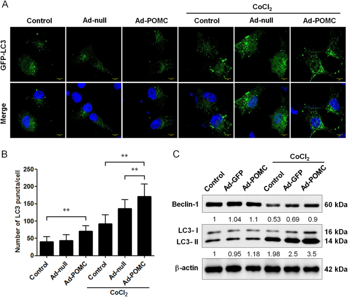
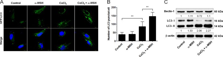
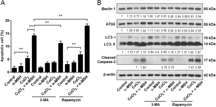
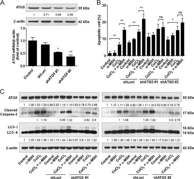
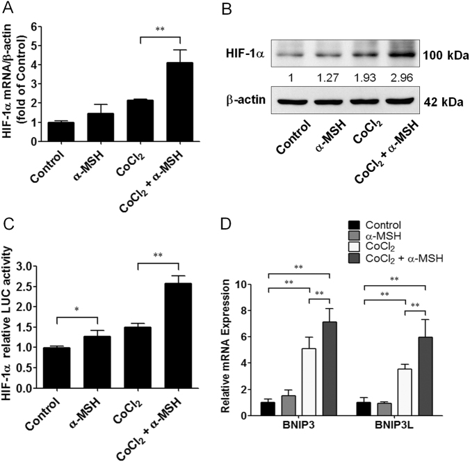
Hypoxia in tumors is known to trigger the pro-survival pathways such as autophagy. Systemic proopiomelanocortin (POMC) gene therapy suppresses melanoma through apoptosis induction and neovascularization blockage. In this study, we investigated the crosstalk between autophagic and apoptotic signaling in POMC-mediated melanoma suppression. By histological and immunoblot analysis, it was shown that POMC-treated melanoma tissues exhibited the prominent LC3 immunostaining, which was correlated with reduced CD31-positive tumor vascularization. Such autophagy induction could be recapitulated in melanoma cells receiving POMC gene delivery and hypoxia-mimicking agent cobalt chloride (CoCl). We then utilized the POMC-derived peptide α-MSH with CoCl to elicit the autophagy as well as apoptosis in cultured melanoma cells. To delineate the role of autophagy during cell death, application of autophagy-inducer rapamycin enhanced, whereas autophagy inhibitor 3-MA attenuated, the α-MSH-induced apoptosis in melanoma cells. Genetic silencing of ATG5, an autophagy regulator, by RNA interference perturbed the α-MSH-induced apoptosis in melanoma cells. Finally, it was delineated that α-MSH stimulated the HIF-1α signaling as well as the expression of BNIP3/BNIP3L, thereby promoting the autophagy and apoptosis in melanoma cells. Therefore, the present study unveiled a unique function of autophagy in promoting cell death during POMC-mediated melanoma suppression via α-MSH/HIF-1α/BNIP3/BNIP3L signaling pathway.
已知肿瘤中的缺氧会触发自噬等促生存途径。系统性阿片-促黑素细胞皮质素(POMC)基因疗法通过诱导凋亡和阻断新血管形成来抑制黑色素瘤。在本研究中,我们调查了POMC介导的黑色素瘤抑制过程中自噬信号与凋亡信号之间的相互作用。通过组织学和免疫印迹分析表明,接受POMC治疗的黑色素瘤组织呈现出显著的LC3免疫染色,这与CD31阳性肿瘤血管生成减少相关。这种自噬诱导在接受POMC基因传递和缺氧模拟剂氯化钴(CoCl)的黑色素瘤细胞中也可重现。然后我们利用POMC衍生肽α-MSH与CoCl在培养的黑色素瘤细胞中引发自噬以及凋亡。为了阐明自噬在细胞死亡过程中的作用,自噬诱导剂雷帕霉素的应用增强了α-MSH诱导的黑色素瘤细胞凋亡,而自噬抑制剂3-MA则减弱了这种凋亡。通过RNA干扰对自噬调节因子ATG5进行基因沉默扰乱了α-MSH诱导的黑色素瘤细胞凋亡。最后,明确了α-MSH刺激HIF-1α信号以及BNIP3/BNIP3L的表达,从而促进黑色素瘤细胞中的自噬和凋亡。因此,本研究揭示了自噬在通过α-MSH/HIF-1α/BNIP3/BNIP3L信号通路介导的POMC介导的黑色素瘤抑制过程中促进细胞死亡的独特功能。