Tsinghua-Peking Center for Life Sciences, Tsinghua University, Beijing 100084, China; Laboratory of Dynamic Immunobiology, Institute for Immunology, Tsinghua University, Beijing 100084, China; Department of Basic Medical Sciences, School of Medicine, Tsinghua University, Beijing 100084, China; School of Life Sciences, Tsinghua University, Beijing 100084, China; Beijing Key Lab for Immunological Research on Chronic Diseases, Tsinghua University, Beijing 100084, China.
Tsinghua-Peking Center for Life Sciences, Tsinghua University, Beijing 100084, China; Laboratory of Dynamic Immunobiology, Institute for Immunology, Tsinghua University, Beijing 100084, China; Department of Basic Medical Sciences, School of Medicine, Tsinghua University, Beijing 100084, China; Beijing Key Lab for Immunological Research on Chronic Diseases, Tsinghua University, Beijing 100084, China.
Immunity. 2018 Aug 21;49(2):264-274.e4. doi: 10.1016/j.immuni.2018.06.012. Epub 2018 Jul 31.
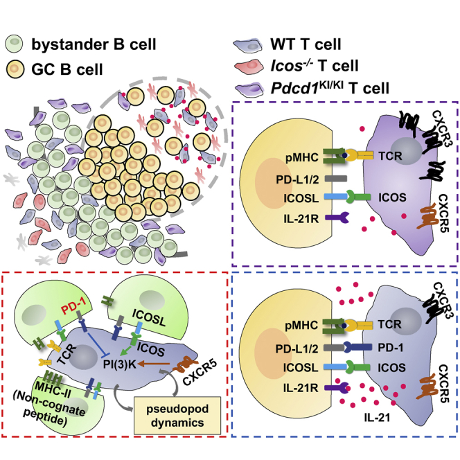
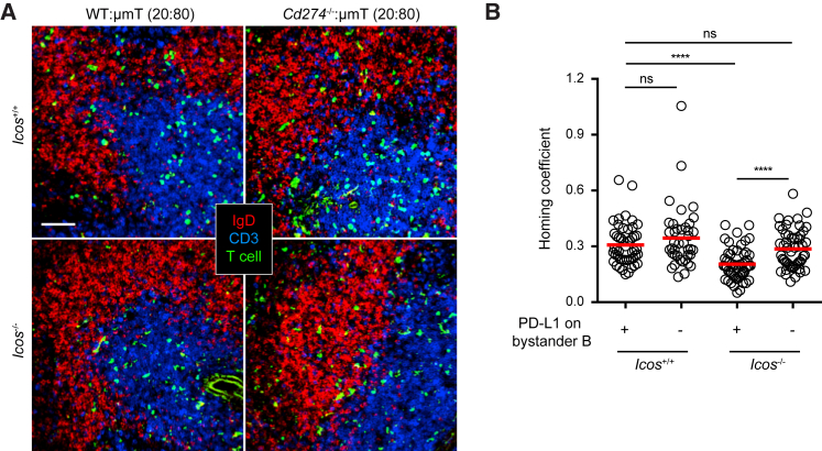
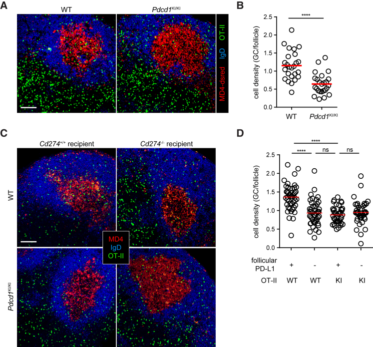
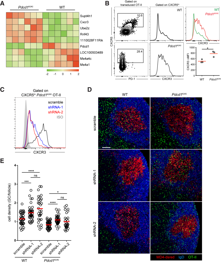
Follicular T helper (Tfh) cells highly express the programmed cell death-1 (PD-1) molecule. Whereas inhibition of T cell receptor (TCR) signaling and CD28 co-stimulation is thought to be the primary mode of PD-1 functions, whether and how PD-1 regulates Tfh cell development and function is unclear. Here we showed that, when engaged by the ensemble of bystander B cells constitutively expressing PD-1 ligand 1 (PD-L1), PD-1 inhibited T cell recruitment into the follicle. This inhibition involved suppression of PI3K activities downstream of the follicle-guidance receptor CXCR5, was independent of co-signaling with the TCR, and necessitated ICOS signaling to overcome. PD-1 further restricted CXCR3 upregulation on Tfh cells, serving to concentrate these cells toward the germinal center territory, where PD-L1-PD-1 interactions between individual Tfh and B cells optimized B cell competition and affinity maturation. Therefore, operating in both costimulation-independent and -dependent manners, PD-1 controls tissue positioning and function of Tfh cells.
滤泡辅助性 T 细胞(Tfh)高度表达程序性细胞死亡受体 1(PD-1)分子。尽管抑制 T 细胞受体(TCR)信号和 CD28 共刺激被认为是 PD-1 功能的主要模式,但 PD-1 是否以及如何调节 Tfh 细胞的发育和功能尚不清楚。在这里,我们发现,当被组成性表达 PD-1 配体 1(PD-L1)的旁观者 B 细胞群体结合时,PD-1 抑制了 T 细胞向滤泡的募集。这种抑制涉及抑制滤泡导向受体 CXCR5 下游的 PI3K 活性,与 TCR 的共信号无关,并且需要 ICOS 信号来克服。PD-1 进一步限制了 Tfh 细胞上 CXCR3 的上调,有助于将这些细胞集中到生发中心区域,在那里个体 Tfh 和 B 细胞之间的 PD-L1-PD-1 相互作用优化了 B 细胞竞争和亲和力成熟。因此,PD-1 以非依赖性和依赖性方式控制 Tfh 细胞的组织定位和功能。