Department of Anesthesiology and Neurology, Shock Trauma and Anesthesiology Research Center, University of Maryland School of Medicine, Baltimore, Maryland.
Department of Neurology, The First Hospital of Jilin University, Changchun, China.
Brain Behav. 2018 Jun;8(6):e00993. doi: 10.1002/brb3.993. Epub 2018 Apr 27.
Neuroprotective therapeutics achieved from animal studies have not been able to translate into clinical stroke therapies. A major reason may be that the functional tests and outcomes between animal stroke studies and clinical trials are significantly different. Ultimately, functional recovery is most important for stroke patients, but it remains challenging to identify animal functional tests that reflect human stroke deficits. This study aimed to explore whether the nest-building activity can be used as a functional test of mouse stroke deficit.
Forty-one C57B6 male mice were randomly assigned into a sham-operated control group and 20-, 40- and 60-min middle cerebral artery occlusion (MCAO) groups. Mice were perfusion-fixed at 21 days following sham surgery or MCAO. Infarct volumes were assessed under the light microscopy. The nest-building activity was characterized and quantitatively evaluated.
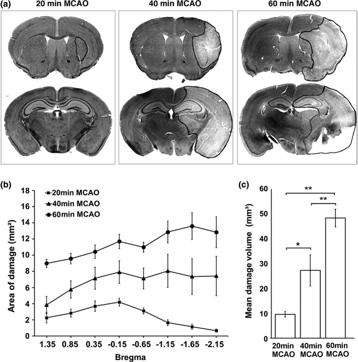
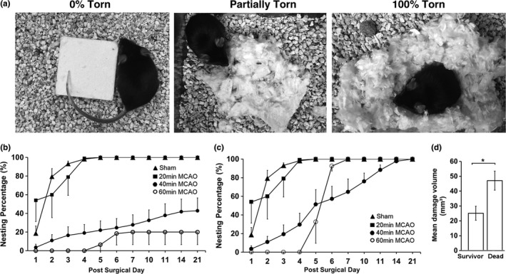
The results show that only a small portion of striatum was damaged after 20-min MCAO. The brain damage areas were expanded from striatum to the neocortex and hippocampus proportionally after 40-min and 60-min MCAO, respectively. Consistently, relative to that of the sham-operated mice, the nest-building activity was insignificantly altered after 20-min MCAO, but dramatically and significantly reduced proportionally following 40-min and 60-min MCAO, respectively. The nest-building deficit was a long-lasting event and could be seen for as long as 14-21 days of recovery, the longest endpoint of this study.
The results suggest that the nest-building activity may be a novel, objective, easy to use, highly sensitive, and long-lasting test that may reflect the multifaceted sensorimotor and cognitive deficits after stroke in humans. Our findings may provide a novel multifaceted test for bridging the gap between animal stroke studies and clinical trials.
从动物研究中获得的神经保护疗法未能转化为临床中风治疗。一个主要原因可能是动物中风研究和临床试验之间的功能测试和结果有很大的不同。最终,对于中风患者来说,功能的恢复是最重要的,但确定反映人类中风缺陷的动物功能测试仍然具有挑战性。本研究旨在探讨筑巢活动是否可以作为小鼠中风缺陷的功能测试。
41 只 C57B6 雄性小鼠被随机分为假手术对照组和 20 分钟、40 分钟和 60 分钟大脑中动脉闭塞(MCAO)组。假手术后或 MCAO 后 21 天,小鼠进行灌注固定。在光镜下评估梗死体积。对筑巢活动进行了特征描述和定量评估。
结果表明,20 分钟 MCAO 后仅有一小部分纹状体受损。40 分钟和 60 分钟 MCAO 后,脑损伤区域从纹状体到新皮质和海马呈比例扩大。与假手术组相比,20 分钟 MCAO 后筑巢活动变化不明显,但 40 分钟和 60 分钟 MCAO 后筑巢活动显著降低。筑巢缺陷是一个持久的事件,在长达 14-21 天的恢复时间内都可以看到,这是本研究的最长终点。
结果表明,筑巢活动可能是一种新颖、客观、易于使用、高度敏感和持久的测试,可以反映人类中风后的多种感觉运动和认知缺陷。我们的研究结果可能为弥合动物中风研究和临床试验之间的差距提供一种新的多方面测试。