Zou Haixiao, Song Li, Jia Mengqi, Wang Li, Sun Yanfang
Department of Stomatology, the Second Affiliated Hospital of Nanchang University, Nanchang State Key Laboratory Breeding Base of Basic Science of Stomatology (Hubei-MOST), Key Laboratory of Oral Biomedicine Ministry of Education Department of Oral and Maxillofacial Surgery Department of Pathology, School and Hospital of Stomatology, Wuhan University, Wuhan, People's Republic of China.
Medicine (Baltimore). 2018 Aug;97(33):e11877. doi: 10.1097/MD.0000000000011877.
Only 4.5% of brown tumors involve facial bones; of these, solitary bone involvement is usual. Brown tumors of multiple facial bones are extremely rare. Here, we report the case of a brown tumor of multiple facial bones initially misdiagnosed as an odontogenic cyst.
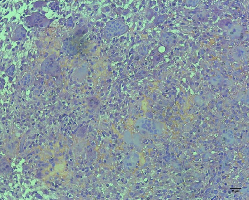
A pregnant 26-year-old woman was referred to our hospital with painful swelling of multiple facial bones, anemia, urinary calculi, marasmus, and a history of multiple bone fractures. Laboratory examination revealed an elevated serum calcium level of 3.09 mmol/L (normal range: 2.0-2.8 mmol/L) and a low phosphorus level of 0.62 mmol/L (normal range: 0.81-1.65 mmol/L). The serum alkaline phosphatase concentration was 397 IU/L (normal range: 24-82 IU/L) and parathyroid hormone level was 267 pg/mL (normal range: 14-72 pg/mL). Cone beam computed tomography revealed multiple ossifying fibromas of the maxilla and mandible. Incisional biopsy revealed abundant spindle cells with areas of hemorrhage and haphazardly arranged diffuse multinucleated giant cells.
The patient was diagnosed with primary hyperparathyroidism (HPT).
She was treated by parathyroidectomy.
The multiple osteitis fibrosa cystica gradually resolved as bone re-mineralized. The patient has been followed up for 2 years without evidence of tumor recurrence.
As multiple osteolytic lesions of facial bones can be caused by primary HPT, serum calcium and parathyroid hormone assays should be performed routinely when investigating these lesions.
棕色瘤仅4.5%累及面骨;其中,通常为单骨受累。多块面骨发生棕色瘤极为罕见。在此,我们报告一例最初被误诊为牙源性囊肿的多块面骨棕色瘤病例。
一名26岁孕妇因多块面骨疼痛性肿胀、贫血、尿路结石、消瘦及多次骨折史被转诊至我院。实验室检查显示血清钙水平升高至3.09 mmol/L(正常范围:2.0 - 2.8 mmol/L),磷水平降低至0.62 mmol/L(正常范围:0.81 - 1.65 mmol/L)。血清碱性磷酸酶浓度为397 IU/L(正常范围:24 - 82 IU/L),甲状旁腺激素水平为267 pg/mL(正常范围:14 - 72 pg/mL)。锥形束计算机断层扫描显示上颌骨和下颌骨有多个骨化纤维瘤。切开活检显示有大量梭形细胞,伴有出血区域和排列杂乱的弥漫性多核巨细胞。
患者被诊断为原发性甲状旁腺功能亢进症(HPT)。
对她进行了甲状旁腺切除术治疗。
随着骨再矿化,多发性纤维性骨炎囊肿逐渐消退。患者已随访2年,无肿瘤复发迹象。
由于面骨的多发性溶骨性病变可能由原发性HPT引起,在对这些病变进行调查时应常规检测血清钙和甲状旁腺激素。