• 文献检索
  • 文档翻译
  • 深度研究
  • 学术资讯
  • Suppr Zotero 插件Zotero 插件
  • 邀请有礼
  • 套餐&价格
  • 历史记录
应用&插件
Suppr Zotero 插件Zotero 插件浏览器插件Mac 客户端Windows 客户端微信小程序
定价
高级版会员购买积分包购买API积分包
服务
文献检索文档翻译深度研究API 文档MCP 服务
关于我们
关于 Suppr公司介绍联系我们用户协议隐私条款
关注我们

Suppr 超能文献

核心技术专利:CN118964589B侵权必究
粤ICP备2023148730 号-1Suppr @ 2026

文献检索

告别复杂PubMed语法,用中文像聊天一样搜索,搜遍4000万医学文献。AI智能推荐,让科研检索更轻松。

立即免费搜索

文件翻译

保留排版,准确专业,支持PDF/Word/PPT等文件格式,支持 12+语言互译。

免费翻译文档

深度研究

AI帮你快速写综述,25分钟生成高质量综述,智能提取关键信息,辅助科研写作。

立即免费体验

2型糖尿病病程中眼表损伤的评估

Assessment of Ocular Surface Damage during the Course of Type 2 Diabetes Mellitus.

作者信息

He Fanglin, Zhao Zhanlin, Liu Yan, Lu Linna, Fu Yao

机构信息

Department of Ophthalmology, Shanghai Ninth People's Hospital, Shanghai Jiaotong University School of Medicine, Shanghai, China.

Shanghai Key Laboratory of Orbital Disease and Ocular Oncology, Shanghai, China.

出版信息

J Ophthalmol. 2018 Jul 15;2018:1206808. doi: 10.1155/2018/1206808. eCollection 2018.

DOI:10.1155/2018/1206808
PMID:30116624
原文链接:https://pmc.ncbi.nlm.nih.gov/articles/PMC6079525/
Abstract

PURPOSE

To investigate the impact of disease duration on the ocular surface during the course of type 2 diabetes mellitus compared with nondiabetic controls.

METHODS

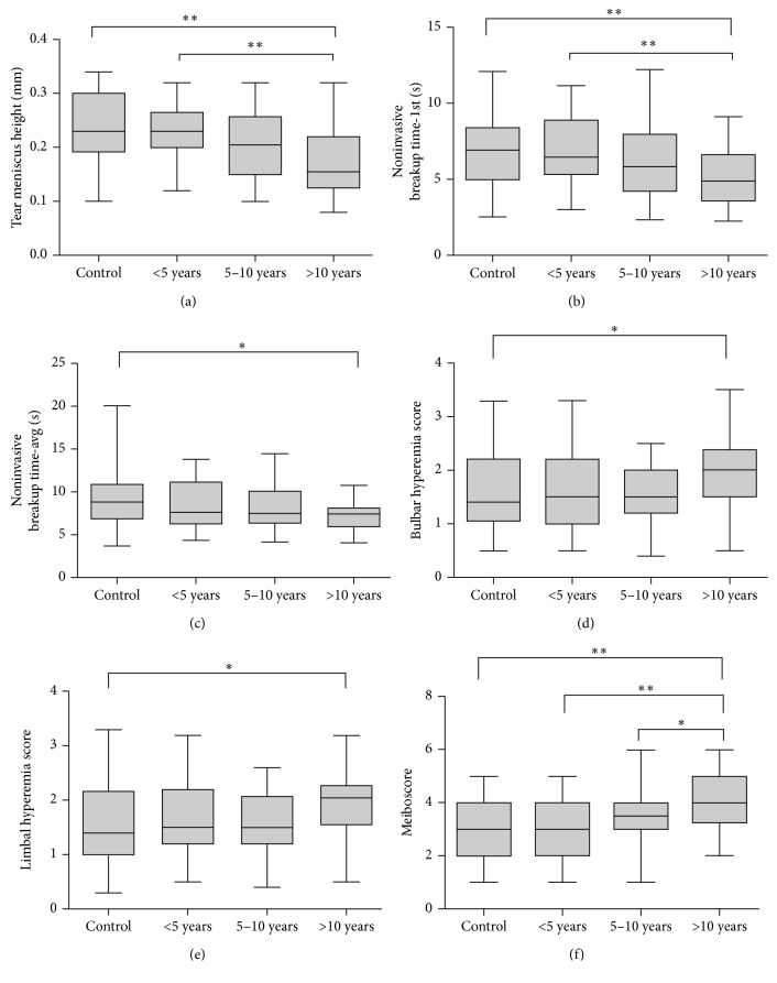
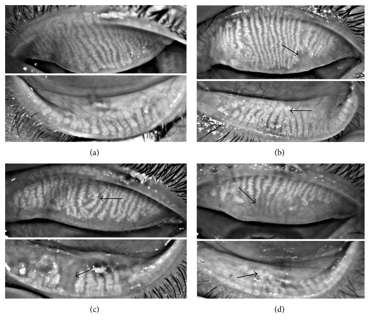
One hundred twenty diabetic patients were divided into three groups according to disease duration: less than 5 years, 5-10 years, and over 10 years. All eyes were imaged using a corneal topographer (Oculus Keratograph 5M). Tear film measurements and meibography were also recorded. Meibomian gland changes were scored from 0 to 6 (meiboscore).

RESULTS

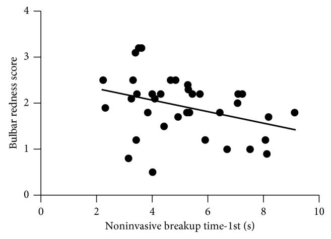
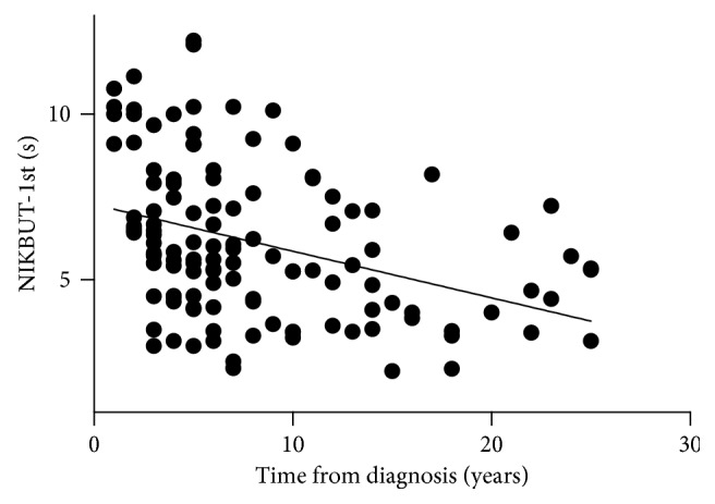
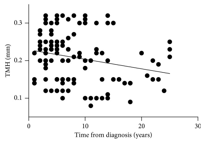
The noninvasive breakup time first (NIKBUT-1st) and noninvasive breakup time average (NIKBUT-avg) were significantly shorter in the over 10 years diabetic group compared with the control group (=0.0056  and  =0.010, resp.). Tear meniscus height (TMH) was significantly lower in the over 10 years diabetic group compared with the control group (=0.0016) and the 5 years group (=0.0061). We also found that more patients in the over 10 years diabetic group showed bulbar and limbal hyperemia compared with the control group (bulbar hyperemia: =0.049; limbal hyperemia: =0.026). The meiboscore in the over 10 years diabetic group was significantly higher compared with the other three groups ( < 0.05). Bulbar hyperemia showed a significant negative correlation with NIKBUT-1st in the over 10 years diabetic group (=-0.35  and   < 0.05).

CONCLUSION

Ocular surface damage in long-term type 2 diabetes is more severe than that in patients with shorter disease duration.

摘要

目的

与非糖尿病对照组相比,研究2型糖尿病病程中疾病持续时间对眼表的影响。

方法

120例糖尿病患者根据病程分为三组:病程小于5年、5至10年、超过10年。所有眼睛均使用角膜地形图仪(Oculus Keratograph 5M)进行成像。还记录了泪膜测量和睑板腺造影。睑板腺变化从0到6分进行评分(睑板腺评分)。

结果

与对照组相比,病程超过10年的糖尿病组首次无创泪膜破裂时间(NIKBUT-1st)和平均无创泪膜破裂时间(NIKBUT-avg)显著缩短(分别为=0.0056和=0.010)。与对照组(=0.0016)和病程5年组(=0.0061)相比,病程超过10年的糖尿病组泪河高度(TMH)显著降低。我们还发现,与对照组相比,病程超过10年的糖尿病组中更多患者出现球结膜和角膜缘充血(球结膜充血:=0.049;角膜缘充血:=0.026)。病程超过10年的糖尿病组睑板腺评分显著高于其他三组(<0.05)。在病程超过10年的糖尿病组中,球结膜充血与NIKBUT-1st呈显著负相关(=-0.35,<0.05)。

结论

长期2型糖尿病患者的眼表损伤比病程较短的患者更严重。

https://cdn.ncbi.nlm.nih.gov/pmc/blobs/2489/6079525/69e3979bbb09/JOPH2018-1206808.005.jpg
https://cdn.ncbi.nlm.nih.gov/pmc/blobs/2489/6079525/4f2799712579/JOPH2018-1206808.001.jpg
https://cdn.ncbi.nlm.nih.gov/pmc/blobs/2489/6079525/ebc1e9d517fd/JOPH2018-1206808.002.jpg
https://cdn.ncbi.nlm.nih.gov/pmc/blobs/2489/6079525/15c98cb52190/JOPH2018-1206808.003.jpg
https://cdn.ncbi.nlm.nih.gov/pmc/blobs/2489/6079525/e67ba86956cb/JOPH2018-1206808.004.jpg
https://cdn.ncbi.nlm.nih.gov/pmc/blobs/2489/6079525/69e3979bbb09/JOPH2018-1206808.005.jpg

相似文献

1
Assessment of Ocular Surface Damage during the Course of Type 2 Diabetes Mellitus.2型糖尿病病程中眼表损伤的评估
J Ophthalmol. 2018 Jul 15;2018:1206808. doi: 10.1155/2018/1206808. eCollection 2018.
2
Alterations of Ocular Surface and Tear Film in Patients with Obstructive Sleep Apnea/Hypopnea Syndrome.阻塞性睡眠呼吸暂停低通气综合征患者眼表及泪膜的改变
Nat Sci Sleep. 2022 Feb 23;14:277-290. doi: 10.2147/NSS.S340105. eCollection 2022.
3
Noninvasive Imaging of Tear Film Dynamics in Eyes With Ocular Surface Disease.眼表疾病患者泪膜动力学的无创成像
Cornea. 2015 Oct;34 Suppl 10:S48-52. doi: 10.1097/ICO.0000000000000570.
4
Meibomian Gland Dysfunction in Type 2 Diabetic Patients.2型糖尿病患者的睑板腺功能障碍
J Ophthalmol. 2017;2017:3047867. doi: 10.1155/2017/3047867. Epub 2017 May 16.
5
Comparison of Each Eye According to the Order of Noninvasive Keratographic Tear Film Evaluation.根据非侵入性角膜泪膜评估的顺序比较每只眼睛。
Korean J Ophthalmol. 2023 Jun;37(3):230-235. doi: 10.3341/kjo.2022.0135. Epub 2023 Jun 5.
6
A Novel Noninvasive Screening Tool for Dry Eye Disease.一种用于干眼症的新型非侵入性筛查工具。
Diagnostics (Basel). 2024 Jun 7;14(12):1209. doi: 10.3390/diagnostics14121209.
7
Keratograph 5M As A Useful And Objective Tool For Evaluating The Ocular Surface In Limbal Stem Cell Deficiency.角膜地形图仪5M作为评估角膜缘干细胞缺乏症眼表的一种有用且客观的工具
Clin Ophthalmol. 2019 Oct 16;13:2025-2033. doi: 10.2147/OPTH.S218313. eCollection 2019.
8
Comparative Evaluation of Clinical Methods of Tear Film Stability Assessment: A Randomized Crossover Trial.比较评估泪膜稳定性评估的临床方法:一项随机交叉试验。
JAMA Ophthalmol. 2018 Mar 1;136(3):291-294. doi: 10.1001/jamaophthalmol.2017.6489.
9
Correlation of the retinopathy degree with the change of ocular surface and corneal nerve in patients with type 2 diabetes mellitus.2型糖尿病患者视网膜病变程度与眼表及角膜神经变化的相关性
Int J Ophthalmol. 2021 May 18;14(5):750-758. doi: 10.18240/ijo.2021.05.17. eCollection 2021.
10
Dry Eye Indexes Estimated by Keratograph 5M of Systemic Lupus Erythematosus Patients without Secondary Sjögren's Syndrome Correlate with Lupus Activity.通过角膜地形图5M评估的无继发性干燥综合征的系统性红斑狼疮患者的干眼指数与狼疮活动相关。
J Ophthalmol. 2019 Aug 29;2019:8509089. doi: 10.1155/2019/8509089. eCollection 2019.

引用本文的文献

1
Characteristics and associated risk factors of exposure keratopathy among ventilated patients in intensive care units in Jordan.约旦重症监护病房中接受机械通气患者暴露性角膜病变的特征及相关危险因素
Acute Crit Care. 2025 Apr 11;40(2):330-8. doi: 10.4266/acc.003648.
2
Meibomian Gland Dysfunction and Dropout in Diabetic Patients with Non-Proliferative Diabetic Retinopathy.非增殖性糖尿病视网膜病变糖尿病患者的睑板腺功能障碍与脱落
Bioengineering (Basel). 2024 Sep 10;11(9):907. doi: 10.3390/bioengineering11090907.
3
Influencing factors of self-management ability among dry eye patients in west China.

本文引用的文献

1
Conjunctival Tear Layer Temperature, Evaporation, Hyperosmolarity, Inflammation, Hyperemia, Tissue Damage, and Symptoms: A Review of an Amplifying Cascade.结膜泪膜温度、蒸发、高渗、炎症、充血、组织损伤及症状:一个放大级联反应的综述
Curr Eye Res. 2017 Dec;42(12):1574-1584. doi: 10.1080/02713683.2017.1377261. Epub 2017 Nov 7.
2
TFOS DEWS II pathophysiology report.TFOS DEWS II 病理生理学报告。
Ocul Surf. 2017 Jul;15(3):438-510. doi: 10.1016/j.jtos.2017.05.011. Epub 2017 Jul 20.
3
TFOS DEWS II Epidemiology Report.TFOS DEWS II 流行病学报告。
中国西部干眼患者自我管理能力的影响因素
Int J Ophthalmol. 2024 Sep 18;17(9):1621-1627. doi: 10.18240/ijo.2024.09.07. eCollection 2024.
4
Immune-Mediated Ocular Surface Disease in Diabetes Mellitus-Clinical Perspectives and Treatment: A Narrative Review.糖尿病中的免疫介导性眼表疾病——临床观点与治疗:一项叙述性综述
Biomedicines. 2024 Jun 12;12(6):1303. doi: 10.3390/biomedicines12061303.
5
Immune Fingerprint in Diabetes: Ocular Surface and Retinal Inflammation.糖尿病的免疫指纹:眼表和视网膜炎症。
Int J Mol Sci. 2023 Jun 6;24(12):9821. doi: 10.3390/ijms24129821.
6
Metabolomics in Corneal Diseases: A Narrative Review from Clinical Aspects.角膜疾病中的代谢组学:从临床角度的叙述性综述
Metabolites. 2023 Mar 3;13(3):380. doi: 10.3390/metabo13030380.
7
Putative Biomarkers in Tears for Diabetic Retinopathy Diagnosis.用于糖尿病视网膜病变诊断的泪液中潜在生物标志物
Front Med (Lausanne). 2022 May 25;9:873483. doi: 10.3389/fmed.2022.873483. eCollection 2022.
8
Ocular Surface Diseases in Patients With Diabetes.糖尿病患者的眼表疾病
Cureus. 2022 Mar 22;14(3):e23401. doi: 10.7759/cureus.23401. eCollection 2022 Mar.
9
Metabolic phenotyping of tear fluid as a prognostic tool for personalised medicine exemplified by T2DM patients.以2型糖尿病患者为例,泪液代谢表型分析作为个性化医疗的预后工具
EPMA J. 2022 Jan 29;13(1):107-123. doi: 10.1007/s13167-022-00272-7. eCollection 2022 Mar.
10
Morphology of Meibomian Glands in a 65-Year-Old Norwegian Population without Dry Eye Disease.65岁无干眼疾病的挪威人群睑板腺的形态学
J Clin Med. 2022 Jan 20;11(3):527. doi: 10.3390/jcm11030527.
Ocul Surf. 2017 Jul;15(3):334-365. doi: 10.1016/j.jtos.2017.05.003. Epub 2017 Jul 20.
4
TFOS DEWS II Definition and Classification Report.TFOS DEWS II 定义与分类报告。
Ocul Surf. 2017 Jul;15(3):276-283. doi: 10.1016/j.jtos.2017.05.008. Epub 2017 Jul 20.
5
Meibomian Gland Dysfunction in Type 2 Diabetic Patients.2型糖尿病患者的睑板腺功能障碍
J Ophthalmol. 2017;2017:3047867. doi: 10.1155/2017/3047867. Epub 2017 May 16.
6
Diabetic retinopathy: global prevalence, major risk factors, screening practices and public health challenges: a review.糖尿病视网膜病变:全球患病率、主要危险因素、筛查实践及公共卫生挑战:一项综述
Clin Exp Ophthalmol. 2016 May;44(4):260-77. doi: 10.1111/ceo.12696. Epub 2016 Feb 17.
7
A Clinical Study of Meibomian Gland Dysfunction in Patients with Diabetes.糖尿病患者睑板腺功能障碍的临床研究
Middle East Afr J Ophthalmol. 2015 Oct-Dec;22(4):462-6. doi: 10.4103/0974-9233.167827.
8
Effects of Insulin and High Glucose on Human Meibomian Gland Epithelial Cells.胰岛素和高糖对人睑板腺上皮细胞的影响。
Invest Ophthalmol Vis Sci. 2015 Dec;56(13):7814-20. doi: 10.1167/iovs.15-18049.
9
Ocular surface disease in patients with diabetic peripheral neuropathy.糖尿病周围神经病变患者的眼表疾病
Br J Ophthalmol. 2016 Jul;100(7):924-928. doi: 10.1136/bjophthalmol-2015-307369. Epub 2015 Oct 23.
10
Noninvasive Imaging of Tear Film Dynamics in Eyes With Ocular Surface Disease.眼表疾病患者泪膜动力学的无创成像
Cornea. 2015 Oct;34 Suppl 10:S48-52. doi: 10.1097/ICO.0000000000000570.