• 文献检索
  • 文档翻译
  • 深度研究
  • 学术资讯
  • Suppr Zotero 插件Zotero 插件
  • 邀请有礼
  • 套餐&价格
  • 历史记录
应用&插件
Suppr Zotero 插件Zotero 插件浏览器插件Mac 客户端Windows 客户端微信小程序
定价
高级版会员购买积分包购买API积分包
服务
文献检索文档翻译深度研究API 文档MCP 服务
关于我们
关于 Suppr公司介绍联系我们用户协议隐私条款
关注我们

Suppr 超能文献

核心技术专利:CN118964589B侵权必究
粤ICP备2023148730 号-1Suppr @ 2026

文献检索

告别复杂PubMed语法,用中文像聊天一样搜索,搜遍4000万医学文献。AI智能推荐,让科研检索更轻松。

立即免费搜索

文件翻译

保留排版,准确专业,支持PDF/Word/PPT等文件格式,支持 12+语言互译。

免费翻译文档

深度研究

AI帮你快速写综述,25分钟生成高质量综述,智能提取关键信息,辅助科研写作。

立即免费体验

使用造影剂和弹性成像技术的超声内镜肝脏评估

EUS liver assessment using contrast agents and elastography.

作者信息

Lisotti Andrea, Serrani Marta, Caletti Giancarlo, Fusaroli Pietro

机构信息

Department of Medical and Surgical Sciences, Gastroenterology Unit, Hospital of Imola, University of Bologna, Bologna, Italy.

出版信息

Endosc Ultrasound. 2018 Jul-Aug;7(4):252-256. doi: 10.4103/eus.eus_29_18.

DOI:10.4103/eus.eus_29_18
PMID:30117488
原文链接:https://pmc.ncbi.nlm.nih.gov/articles/PMC6106147/
Abstract

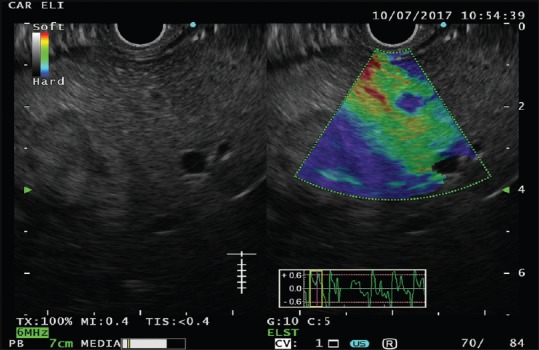
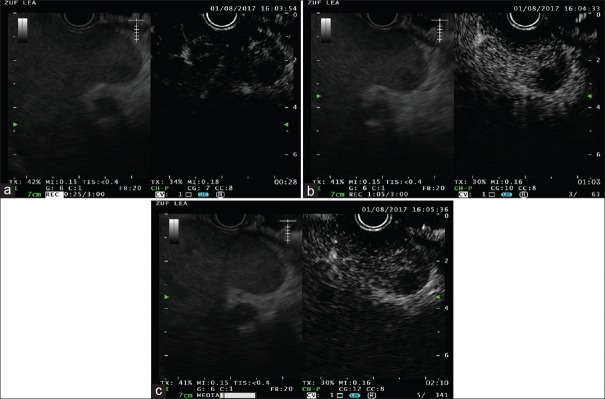
Transabdominal-US is the first-line imaging modality used to assess the whole liver parenchyma and vascularization; EUS assessment of the liver is incomplete and is not sufficient to rule out the presence of focal liver lesions. On the other hand, due the high diagnostic yield in detecting very small (< 1 cm) lesions, EUS is considered complementary to radiological imaging techniques for the investigation of liver parenchyma. Scarce data are available regarding the investigation of liver parenchyma using both EUS-elastography (EUS-E) and CH-EUS. The aim of this review is to evaluate the clinical role of image enhancement techniques, namely EUS-E and contrast harmonic-EUS (CH-EUS), for the evaluation liver diseases. Despite a potential interest for the application of EUS-E in the assessment of liver diseases, available evidence relegates this technique only to research areas, such as the differential diagnosis between benign and malignant focal liver lesions and the quantification of liver fibrosis in diffuse parenchymal diseases. With the future introduction of EUS shear-wave elastography, interesting data can be obtained for the assessment of liver fibrosis during real-time EUS evaluation. The usefulness of CH-EUS for the evaluation of liver disease is limited by the intrinsic EUS ability to explore only the left lobe and a small part of the right lobe. CH-EUS could be used to increase the diagnostic ability of EUS for the detection and characterization of small lesions and for guiding tissue sampling. Targeting EUS-guided treatments with either EUS-E or CH-EUS might represent potential future applications.

摘要

经腹超声是用于评估整个肝实质和血管化的一线成像方式;超声内镜对肝脏的评估不完整,不足以排除肝脏局灶性病变的存在。另一方面,由于超声内镜在检测非常小(<1cm)的病变方面具有较高的诊断率,因此在肝实质检查中被认为是放射成像技术的补充。关于同时使用超声内镜弹性成像(EUS-E)和对比谐波超声内镜(CH-EUS)对肝实质进行检查的数据较少。本综述的目的是评估图像增强技术,即EUS-E和对比谐波超声内镜(CH-EUS)在评估肝脏疾病中的临床作用。尽管EUS-E在肝脏疾病评估中的应用可能具有一定价值,但现有证据仅将该技术局限于研究领域,如肝脏局灶性病变的良恶性鉴别诊断以及弥漫性实质疾病中肝纤维化的量化。随着超声内镜剪切波弹性成像技术的未来引入,在实时超声内镜评估过程中可获得有关肝纤维化评估的有趣数据。CH-EUS对肝脏疾病评估的有用性受到超声内镜本身仅能探查左叶和右叶一小部分的限制。CH-EUS可用于提高超声内镜对小病变的检测和特征描述能力以及引导组织采样。使用EUS-E或CH-EUS靶向超声内镜引导治疗可能代表未来的潜在应用。

https://cdn.ncbi.nlm.nih.gov/pmc/blobs/a8d1/6106147/96e63e9e005e/EUS-7-252-g003.jpg
https://cdn.ncbi.nlm.nih.gov/pmc/blobs/a8d1/6106147/34909fa99ba0/EUS-7-252-g001.jpg
https://cdn.ncbi.nlm.nih.gov/pmc/blobs/a8d1/6106147/cdb1cccd4ce4/EUS-7-252-g002.jpg
https://cdn.ncbi.nlm.nih.gov/pmc/blobs/a8d1/6106147/96e63e9e005e/EUS-7-252-g003.jpg
https://cdn.ncbi.nlm.nih.gov/pmc/blobs/a8d1/6106147/34909fa99ba0/EUS-7-252-g001.jpg
https://cdn.ncbi.nlm.nih.gov/pmc/blobs/a8d1/6106147/cdb1cccd4ce4/EUS-7-252-g002.jpg
https://cdn.ncbi.nlm.nih.gov/pmc/blobs/a8d1/6106147/96e63e9e005e/EUS-7-252-g003.jpg

相似文献

1
EUS liver assessment using contrast agents and elastography.使用造影剂和弹性成像技术的超声内镜肝脏评估
Endosc Ultrasound. 2018 Jul-Aug;7(4):252-256. doi: 10.4103/eus.eus_29_18.
2
Ultrasound Shear Wave Elastography for Liver Disease. A Critical Appraisal of the Many Actors on the Stage.用于肝脏疾病的超声剪切波弹性成像:对该领域众多参与者的批判性评估
Ultraschall Med. 2016 Feb;37(1):1-5. doi: 10.1055/s-0035-1567037. Epub 2016 Feb 12.
3
Role of contrast-enhanced harmonic endoscopic ultrasonography (EUS) and EUS elastography in pancreatic lesions.对比增强谐波内镜超声检查(EUS)及EUS弹性成像在胰腺病变中的作用
Clin Endosc. 2024 Mar;57(2):164-174. doi: 10.5946/ce.2023.074. Epub 2024 Jan 17.
4
Value of image enhancement of endoscopic ultrasound for diagnosis of gastrointestinal subepithelial lesions.内镜超声图像增强在胃肠道上皮下病变诊断中的价值
DEN Open. 2024 Oct 12;5(1):e70026. doi: 10.1002/deo2.70026. eCollection 2025 Apr.
5
Contrast enhancement and elastography in endoscopic ultrasound: an update of clinical applications in pancreatic diseases.内镜超声中的对比增强与弹性成像:胰腺疾病临床应用的最新进展
Minerva Med. 2016 Aug;107(4):217-22. Epub 2016 May 17.
6
Improved detection of liver metastasis using Kupffer-phase imaging in contrast-enhanced harmonic EUS in patients with pancreatic cancer (with video).应用超声造影谐波 Kupffer 期成像增强超声内镜对胰腺癌患者肝转移的检测(附视频)
Gastrointest Endosc. 2021 Feb;93(2):433-441. doi: 10.1016/j.gie.2020.06.051. Epub 2020 Jun 24.
7
Advances in endoscopic ultrasound imaging of colorectal diseases.结直肠疾病的内镜超声成像进展。
World J Gastroenterol. 2016 Feb 7;22(5):1756-66. doi: 10.3748/wjg.v22.i5.1756.
8
Characterization of small solid tumors in the pancreas: the value of contrast-enhanced harmonic endoscopic ultrasonography.胰腺小实体瘤的特征:对比增强谐波超声内镜检查的价值。
Am J Gastroenterol. 2012 Feb;107(2):303-10. doi: 10.1038/ajg.2011.354. Epub 2011 Oct 18.
9
Current status of tissue harmonic imaging in endoscopic ultrasonography (EUS) and EUS-elastography in pancreatobiliary diseases.组织谐波成像在内镜超声检查(EUS)中的现状以及EUS弹性成像在胰腺疾病中的应用
Dig Endosc. 2015 Apr;27 Suppl 1:68-73. doi: 10.1111/den.12433.
10
Clinical impact of strain histogram EUS elastography and contrast-enhanced EUS for the differential diagnosis of focal pancreatic masses: A prospective multicentric study.应变直方图超声内镜弹性成像和对比增强超声内镜在胰腺局灶性肿块鉴别诊断中的临床应用:一项前瞻性多中心研究。
Endosc Ultrasound. 2020 Mar-Apr;9(2):116-121. doi: 10.4103/eus.eus_69_19.

引用本文的文献

1
Advances in Endo-Hepatology: The Role of Endoscopic Ultrasound in the Management of Portal Hypertension.内镜肝脏病学进展:内镜超声在门静脉高压管理中的作用
Diagnostics (Basel). 2025 Apr 10;15(8):967. doi: 10.3390/diagnostics15080967.
2
The application of artificial intelligence in EUS.人工智能在超声内镜检查中的应用。
Endosc Ultrasound. 2024 Mar-Apr;13(2):65-75. doi: 10.1097/eus.0000000000000053. Epub 2024 Apr 10.
3
Endohepatology: The endoscopic armamentarium in the hand of the hepatologist.内科学:肝病学家手中的内镜设备。

本文引用的文献

1
Improved detection of liver metastasis using Kupffer-phase imaging in contrast-enhanced harmonic EUS in patients with pancreatic cancer (with video).应用超声造影谐波 Kupffer 期成像增强超声内镜对胰腺癌患者肝转移的检测(附视频)
Gastrointest Endosc. 2021 Feb;93(2):433-441. doi: 10.1016/j.gie.2020.06.051. Epub 2020 Jun 24.
2
Contrast-enhanced harmonic endoscopic ultrasonography for differential diagnosis of localized gallbladder lesions.对比增强谐波内镜超声检查在局部胆囊病变鉴别诊断中的应用。
Dig Endosc. 2018 Jan;30(1):98-106. doi: 10.1111/den.12900. Epub 2017 Jul 16.
3
EUS-guided fine-needle core liver biopsy sampling using a novel 19-gauge needle with modified 1-pass, 1 actuation wet suction technique.
Saudi J Gastroenterol. 2024 Jan 1;30(1):4-13. doi: 10.4103/sjg.sjg_214_23. Epub 2023 Nov 18.
4
Endo-hepatology: The changing paradigm of endoscopic ultrasound in cirrhosis.内镜肝脏病学:肝硬化中内镜超声的范式转变
Clin Liver Dis (Hoboken). 2022 Dec 12;20(6):209-215. doi: 10.1002/cld.1263. eCollection 2022 Dec.
5
The usefulness of endoscopic ultrasound in the diagnosis of gallbladder lesions.内镜超声在胆囊病变诊断中的应用价值。
Front Med (Lausanne). 2022 Aug 29;9:957557. doi: 10.3389/fmed.2022.957557. eCollection 2022.
6
Advanced EUS Imaging Techniques.先进的超声内镜成像技术
Dig Dis Sci. 2022 May;67(5):1588-1598. doi: 10.1007/s10620-022-07486-9. Epub 2022 Apr 22.
7
Role of Endoscopic Ultrasound in Liver Disease: Where Do We Stand?内镜超声在肝脏疾病中的作用:我们目前的进展如何?
Diagnostics (Basel). 2021 Oct 31;11(11):2021. doi: 10.3390/diagnostics11112021.
8
The overview of the deep learning integrated into the medical imaging of liver: a review.深度学习在肝脏医学成像中的应用概述:综述
Hepatol Int. 2021 Aug;15(4):868-880. doi: 10.1007/s12072-021-10229-z. Epub 2021 Jul 15.
9
The Role of Interventional Endoscopic Ultrasound in Liver Diseases: What Have We Learnt?介入性内镜超声在肝脏疾病中的作用:我们学到了什么?
Can J Gastroenterol Hepatol. 2021 Jun 28;2021:9948979. doi: 10.1155/2021/9948979. eCollection 2021.
10
Prognostic factors and therapeutic effects of different treatment modalities for colorectal cancer liver metastases.结直肠癌肝转移不同治疗方式的预后因素及治疗效果
World J Gastrointest Oncol. 2020 Oct 15;12(10):1177-1194. doi: 10.4251/wjgo.v12.i10.1177.
EUS 引导下使用新型 19 号针和改良的 1 次 1 触发湿式抽吸技术进行的细针核心肝活检采样。
Gastrointest Endosc. 2018 Feb;87(2):469-475. doi: 10.1016/j.gie.2017.05.013. Epub 2017 May 24.
4
Indications, results, and clinical impact of endoscopic ultrasound (EUS)-guided sampling in gastroenterology: European Society of Gastrointestinal Endoscopy (ESGE) Clinical Guideline - Updated January 2017.内镜超声(EUS)引导下采样在胃肠病学中的适应证、结果及临床影响:欧洲胃肠内镜学会(ESGE)临床指南 - 2017年1月更新版
Endoscopy. 2017 Jul;49(7):695-714. doi: 10.1055/s-0043-109021. Epub 2017 May 16.
5
Contrast harmonic endoscopic ultrasound: Instrumentation, echoprocessors, and echoendoscopes.对比谐波内镜超声:设备、超声处理器和超声内镜
Endosc Ultrasound. 2017 Jan-Feb;6(1):37-42. doi: 10.4103/2303-9027.200214.
6
Harmonic contrast-enhanced endoscopic ultrasound fine-needle aspiration: Fact or fiction?谐波对比增强内镜超声细针穿刺活检:是事实还是虚构?
Endosc Ultrasound. 2017 Jan-Feb;6(1):31-36. doi: 10.4103/2303-9027.196917.
7
Applications of contrast-enhanced harmonic endoscopic ultrasound on biliary, focal liver lesions and vascular diseases.对比增强谐波内镜超声在胆道、肝脏局灶性病变及血管疾病中的应用。
Endosc Ultrasound. 2017 Jan-Feb;6(1):21-24. doi: 10.4103/2303-9027.200211.
8
Dynamic contrast-enhanced endoscopic ultrasound: A quantification method.动态对比增强内镜超声:一种量化方法。
Endosc Ultrasound. 2017 Jan-Feb;6(1):12-20. doi: 10.4103/2303-9027.193595.
9
Maximizing the endosonography: The role of contrast harmonics, elastography and confocal endomicroscopy.最大化内镜超声检查:对比谐波、弹性成像和共聚焦内镜显微镜检查的作用。
World J Gastroenterol. 2017 Jan 7;23(1):25-41. doi: 10.3748/wjg.v23.i1.25.
10
Evaluation of the viability of hepatocellular carcinoma in the caudate lobe using contrast-enhanced endoscopic ultrasonography after transarterial chemoembolization.经动脉化疗栓塞术后使用对比增强内镜超声评估尾状叶肝细胞癌的存活情况。
Endosc Ultrasound. 2016 Nov-Dec;5(6):390-392. doi: 10.4103/2303-9027.190924.