• 文献检索
  • 文档翻译
  • 深度研究
  • 学术资讯
  • Suppr Zotero 插件Zotero 插件
  • 邀请有礼
  • 套餐&价格
  • 历史记录
应用&插件
Suppr Zotero 插件Zotero 插件浏览器插件Mac 客户端Windows 客户端微信小程序
定价
高级版会员购买积分包购买API积分包
服务
文献检索文档翻译深度研究API 文档MCP 服务
关于我们
关于 Suppr公司介绍联系我们用户协议隐私条款
关注我们

Suppr 超能文献

核心技术专利:CN118964589B侵权必究
粤ICP备2023148730 号-1Suppr @ 2026

文献检索

告别复杂PubMed语法,用中文像聊天一样搜索,搜遍4000万医学文献。AI智能推荐,让科研检索更轻松。

立即免费搜索

文件翻译

保留排版,准确专业,支持PDF/Word/PPT等文件格式,支持 12+语言互译。

免费翻译文档

深度研究

AI帮你快速写综述,25分钟生成高质量综述,智能提取关键信息,辅助科研写作。

立即免费体验

对比增强谐波内镜超声在胆道、肝脏局灶性病变及血管疾病中的应用。

Applications of contrast-enhanced harmonic endoscopic ultrasound on biliary, focal liver lesions and vascular diseases.

作者信息

Choi Jun-Ho, Seo Dong-Wan

机构信息

Department of Internal Medicine, Dankook University College of Medicine, Dankook University Hospital, Seoul, South Korea.

Department of Internal Medicine, Division of Gastroenterology, Asan Medical Center, University of Ulsan College of Medicine, Seoul, South Korea.

出版信息

Endosc Ultrasound. 2017 Jan-Feb;6(1):21-24. doi: 10.4103/2303-9027.200211.

DOI:10.4103/2303-9027.200211
PMID:28218196
原文链接:https://pmc.ncbi.nlm.nih.gov/articles/PMC5331838/
Abstract

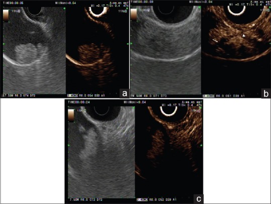
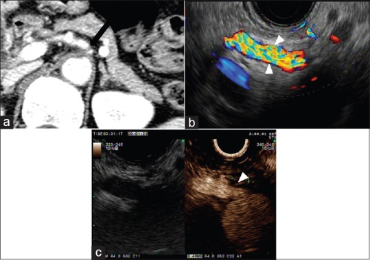
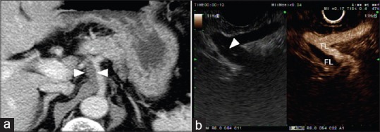
Over the last decade, the clinical applications of contrast-enhanced harmonic endoscopic ultrasound (CH-EUS) have increased steadily. The development of second-generation ultrasound contrast agents has allowed superior visualization of the microvasculature and tissue perfusion of the target lesion. This methodology has proven useful in the differential diagnosis of solid pancreatic masses and lymph nodes. In addition, the applicability of CH-EUS has expanded to nonpancreas structures such as biliary, focal liver lesions, and vascular disease. This article focuses primarily on the novel applications of CH-EUS in biliary tract and visceral vascular diseases.

摘要

在过去十年中,对比增强谐波内镜超声(CH-EUS)的临床应用稳步增加。第二代超声造影剂的发展使得对目标病变的微血管和组织灌注有了更好的可视化效果。这种方法已被证明在胰腺实性肿块和淋巴结的鉴别诊断中很有用。此外,CH-EUS的适用性已扩展到非胰腺结构,如胆道、肝脏局灶性病变和血管疾病。本文主要关注CH-EUS在胆道和内脏血管疾病中的新应用。

https://cdn.ncbi.nlm.nih.gov/pmc/blobs/9eb1/5331838/b1eb5537d50b/EUS-6-21-g003.jpg
https://cdn.ncbi.nlm.nih.gov/pmc/blobs/9eb1/5331838/17421664e9d3/EUS-6-21-g001.jpg
https://cdn.ncbi.nlm.nih.gov/pmc/blobs/9eb1/5331838/e1f94d156d28/EUS-6-21-g002.jpg
https://cdn.ncbi.nlm.nih.gov/pmc/blobs/9eb1/5331838/b1eb5537d50b/EUS-6-21-g003.jpg
https://cdn.ncbi.nlm.nih.gov/pmc/blobs/9eb1/5331838/17421664e9d3/EUS-6-21-g001.jpg
https://cdn.ncbi.nlm.nih.gov/pmc/blobs/9eb1/5331838/e1f94d156d28/EUS-6-21-g002.jpg
https://cdn.ncbi.nlm.nih.gov/pmc/blobs/9eb1/5331838/b1eb5537d50b/EUS-6-21-g003.jpg

相似文献

1
Applications of contrast-enhanced harmonic endoscopic ultrasound on biliary, focal liver lesions and vascular diseases.对比增强谐波内镜超声在胆道、肝脏局灶性病变及血管疾病中的应用。
Endosc Ultrasound. 2017 Jan-Feb;6(1):21-24. doi: 10.4103/2303-9027.200211.
2
Contrast-enhanced harmonic endoscopic ultrasonography for pancreatobiliary diseases.对比增强谐波内镜超声检查在胰腺胆道疾病中的应用
Dig Endosc. 2015 Apr;27 Suppl 1:60-7. doi: 10.1111/den.12454.
3
Contrast enhancement and elastography in endoscopic ultrasound: an update of clinical applications in pancreatic diseases.内镜超声中的对比增强与弹性成像:胰腺疾病临床应用的最新进展
Minerva Med. 2016 Aug;107(4):217-22. Epub 2016 May 17.
4
Contrast-enhanced endoscopic ultrasound.增强型内镜超声检查。
Dig Endosc. 2014 Jan;26 Suppl 1:79-85. doi: 10.1111/den.12179. Epub 2013 Sep 30.
5
Contrast-enhanced harmonic endoscopic ultrasound imaging: basic principles, present situation and future perspectives.对比增强谐波内镜超声成像:基本原理、现状与未来展望。
World J Gastroenterol. 2014 Nov 14;20(42):15549-63. doi: 10.3748/wjg.v20.i42.15549.
6
Contrast-enhanced harmonic endoscopic ultrasonography in pancreatic diseases.对比增强谐波内镜超声检查在胰腺疾病中的应用
Diagn Ther Endosc. 2012;2012:786239. doi: 10.1155/2012/786239. Epub 2012 Nov 1.
7
Characterization of small solid tumors in the pancreas: the value of contrast-enhanced harmonic endoscopic ultrasonography.胰腺小实体瘤的特征:对比增强谐波超声内镜检查的价值。
Am J Gastroenterol. 2012 Feb;107(2):303-10. doi: 10.1038/ajg.2011.354. Epub 2011 Oct 18.
8
Contrast-enhanced harmonic endoscopic ultrasonography for assessment of lymph node metastases in pancreatobiliary carcinoma.对比增强谐波内镜超声检查在胰胆管癌淋巴结转移评估中的应用
World J Gastroenterol. 2016 Mar 28;22(12):3381-91. doi: 10.3748/wjg.v22.i12.3381.
9
Harmonic Contrast-Enhanced Endoscopic Ultrasonography for the Guidance of Fine-Needle Aspiration in Solid Pancreatic Masses.谐波对比增强内镜超声引导下细针穿刺活检实性胰腺肿块的应用
Ultraschall Med. 2017 Apr;38(2):174-182. doi: 10.1055/s-0035-1553496. Epub 2015 Aug 14.
10
Endoscopic ultrasound elastography: Current status and future perspectives.内镜超声弹性成像:现状与未来展望。
World J Gastroenterol. 2015 Dec 21;21(47):13212-24. doi: 10.3748/wjg.v21.i47.13212.

引用本文的文献

1
Utility of Contrast-Enhanced Harmonic Endoscopic Ultrasound for the Guidance and Monitoring of Endoscopic Radiofrequency Ablation.对比增强谐波内镜超声在指导和监测内镜射频消融中的应用。
Gut Liver. 2020 Nov 15;14(6):826-832. doi: 10.5009/gnl19123.
2
Transcutaneous partial pressure of carbon dioxide monitoring during EUS-guided drainage of peripancreatic fluid collections using carbon dioxide insufflation: A prospective study.在使用二氧化碳注入进行内镜超声引导下胰周液体聚集引流期间的经皮二氧化碳分压监测:一项前瞻性研究。
Endosc Ultrasound. 2020 Jan-Feb;9(1):59-65. doi: 10.4103/eus.eus_32_19.
3
EUS liver assessment using contrast agents and elastography.

本文引用的文献

1
The Expanding Role of Contrast-Enhanced Endoscopic Ultrasound in Pancreatobiliary Disease.对比增强内镜超声在胰胆疾病中不断扩大的作用
Gut Liver. 2015 Nov 23;9(6):707-13. doi: 10.5009/gnl15077.
2
Endoscopic ultrasonography-guided ethanol ablation for small pancreatic neuroendocrine tumors: results of a pilot study.内镜超声引导下乙醇消融治疗小胰腺神经内分泌肿瘤:一项初步研究的结果
Clin Endosc. 2015 Mar;48(2):158-64. doi: 10.5946/ce.2015.48.2.158. Epub 2015 Mar 27.
3
Contrast-enhanced harmonic endoscopic ultrasonography in the differential diagnosis of gallbladder wall thickening.
使用造影剂和弹性成像技术的超声内镜肝脏评估
Endosc Ultrasound. 2018 Jul-Aug;7(4):252-256. doi: 10.4103/eus.eus_29_18.
4
Quantitative assessment of hepatic fibrosis in chronic hepatitis B and C: T1 mapping on Gd-EOB-DTPA-enhanced liver magnetic resonance imaging.慢性乙型和丙型肝炎肝纤维化的定量评估:钆塞酸二钠增强肝脏磁共振 T1 mapping 成像。
World J Gastroenterol. 2018 May 14;24(18):2024-2035. doi: 10.3748/wjg.v24.i18.2024.
5
Role of endoscopic ultrasound in liver disease: Where do we stand in 2017?内镜超声在肝脏疾病中的作用:2017年我们处于什么水平?
World J Hepatol. 2017 Aug 28;9(24):1013-1021. doi: 10.4254/wjh.v9.i24.1013.
6
The endoscopist and malignant and non-malignant biliary obstruction.内镜医师与良恶性胆道梗阻。
Biochim Biophys Acta Mol Basis Dis. 2018 Apr;1864(4 Pt B):1478-1483. doi: 10.1016/j.bbadis.2017.09.013. Epub 2017 Sep 18.
对比增强谐波内镜超声检查在胆囊壁增厚鉴别诊断中的应用。
Dig Dis Sci. 2014 Aug;59(8):1909-16. doi: 10.1007/s10620-014-3115-5. Epub 2014 Mar 25.
4
Clinical usefulness with the combination of color Doppler and contrast-enhanced harmonic EUS for the assessment of visceral vascular diseases.彩色多普勒与超声内镜造影谐波成像联合应用于内脏血管疾病评估的临床实用性。
J Clin Gastroenterol. 2014 Nov-Dec;48(10):845-50. doi: 10.1097/MCG.0000000000000032.
5
Utility of contrast-enhanced harmonic EUS in the diagnosis of malignant gallbladder polyps (with videos).超声造影谐波成像在诊断恶性胆囊息肉中的应用(附视频)。
Gastrointest Endosc. 2013 Sep;78(3):484-93. doi: 10.1016/j.gie.2013.03.1328. Epub 2013 Apr 30.
6
Differential diagnosis between gallbladder adenomas and cholesterol polyps on contrast-enhanced harmonic endoscopic ultrasonography.在对比增强谐波内镜超声检查中鉴别胆囊腺瘤和胆固醇息肉。
Surg Endosc. 2013 Apr;27(4):1414-21. doi: 10.1007/s00464-012-2620-x. Epub 2012 Dec 12.
7
Contrast-enhanced ultrasound: The evolving applications.超声造影:不断发展的应用
World J Radiol. 2009 Dec 31;1(1):15-24. doi: 10.4329/wjr.v1.i1.15.
8
Contrast-enhanced harmonic EUS with novel ultrasonographic contrast (Sonazoid) in the preoperative T-staging for pancreaticobiliary malignancies.新型超声造影剂(声诺维)增强谐波内镜超声在胰胆恶性肿瘤术前T分期中的应用
Scand J Gastroenterol. 2010 Jun;45(6):732-8. doi: 10.3109/00365521003690269.
9
Differential diagnosis between benign and malignant gallbladder diseases with real-time contrast-enhanced ultrasound.实时超声造影在胆囊良恶性疾病鉴别诊断中的应用
Eur Radiol. 2010 Jan;20(1):239-48. doi: 10.1007/s00330-009-1538-8. Epub 2009 Aug 6.
10
The utility of contrast-enhanced endoscopic ultrasound in monitoring ethanol-induced pancreatic tissue ablation: a pilot study in a porcine model.对比增强内镜超声在监测乙醇诱导的胰腺组织消融中的应用:一项猪模型的初步研究。
Endoscopy. 2007 Jun;39(6):525-9. doi: 10.1055/s-2007-966391.