Institute of Psychology, Leiden University, Leiden, The Netherlands.
Department of Psychiatry, Leiden University Medical Center, Leiden, The Netherlands.
Neuroimage Clin. 2017 Aug 30;16:678-688. doi: 10.1016/j.nicl.2017.08.001. eCollection 2017.
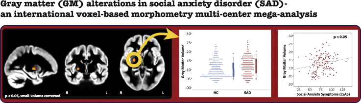
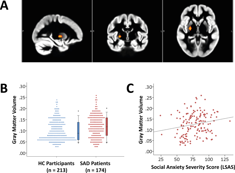
Social anxiety disorder (SAD) is a prevalent and disabling mental disorder, associated with significant psychiatric co-morbidity. Previous research on structural brain alterations associated with SAD has yielded inconsistent results concerning the direction of the changes in gray matter (GM) in various brain regions, as well as on the relationship between brain structure and SAD-symptomatology. These heterogeneous findings are possibly due to limited sample sizes. Multi-site imaging offers new opportunities to investigate SAD-related alterations in brain structure in larger samples. An international multi-center mega-analysis on the largest database of SAD structural T1-weighted 3T MRI scans to date was performed to compare GM volume of SAD-patients ( = 174) and healthy control (HC)-participants ( = 213) using voxel-based morphometry. A hypothesis-driven region of interest (ROI) approach was used, focusing on the basal ganglia, the amygdala-hippocampal complex, the prefrontal cortex, and the parietal cortex. SAD-patients had larger GM volume in the dorsal striatum when compared to HC-participants. This increase correlated positively with the severity of self-reported social anxiety symptoms. No SAD-related differences in GM volume were present in the other ROIs. Thereby, the results of this mega-analysis suggest a role for the dorsal striatum in SAD, but previously reported SAD-related changes in GM in the amygdala, hippocampus, precuneus, prefrontal cortex and parietal regions were not replicated. Our findings emphasize the importance of large sample imaging studies and the need for meta-analyses like those performed by the Enhancing NeuroImaging Genetics through Meta-Analysis (ENIGMA) Consortium.
社交焦虑障碍(SAD)是一种普遍且使人丧失能力的精神障碍,与显著的精神共病有关。以前的研究表明,与 SAD 相关的结构脑改变与灰质(GM)在各个脑区的变化方向以及脑结构与 SAD 症状之间的关系有关,结果不一致。这些异质的发现可能是由于样本量有限。多地点成像为在更大的样本中研究与 SAD 相关的结构脑改变提供了新的机会。对迄今为止最大的 SAD 结构 T1 加权 3T MRI 扫描数据库进行了一项国际多中心 mega 分析,以比较 SAD 患者(n=174)和健康对照组(n=213)的 GM 体积,使用基于体素的形态测量法。使用基于体素的形态测量法,采用了一种基于假设的感兴趣区域(ROI)方法,重点关注基底节、杏仁核-海马复合体、前额叶皮层和顶叶皮层。与 HC 参与者相比,SAD 患者的背侧纹状体 GM 体积更大。这种增加与自我报告的社交焦虑症状的严重程度呈正相关。在其他 ROI 中,GM 体积没有 SAD 相关的差异。因此,这项 mega 分析的结果表明,背侧纹状体在 SAD 中起作用,但以前报道的 SAD 相关 GM 变化在杏仁核、海马体、楔前叶、前额叶和顶叶区域没有得到复制。我们的研究结果强调了大样本成像研究的重要性,以及需要像增强神经成像遗传学通过荟萃分析(ENIGMA)联盟进行的荟萃分析。